Figure 1. Microarray assay experimental design. For microarray analysis 1 (A), RNA from three separate normal (n=3) and three separate POAG (n=3) donor LC cell lines were pooled to individual 'normal'
and 'POAG' microarrays respectively. In microarray analysis 1 LC cells from the normal and POAG donors were passaged in a
1:1 ratio. For microarray analysis 2 (B) a fourth normal and a fourth POAG donor provided RNA which was pooled to a second ‘normal’ and a second ‘POAG’ microarray
respectively. In microarray analysis 2 LC cells from the normal and POAG donors were passaged in a 1:3 ratio. All four arrays
were then normalized together and compared using Robust Multichip Average (RMA) software. The LC cells used were at passages
5 and 6. NLC=normal lamina cribrosa, GLC=POAG lamina cribrosa.
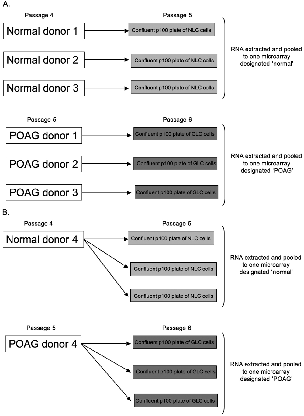
 Figure 1 of
Kirwan, Mol Vis 2009; 15:76-88.
Figure 1 of
Kirwan, Mol Vis 2009; 15:76-88.