Figure 9. Western blot analyses of the expression levels of the p-ERK 1/2 protein in the corneal endothelial cells at various ages of
the SAM R1 and SAM P8 strains. A: The expressions of p-Erk1/2 and Erk1/2 proteins in 1-month-, 6-month-, and 12-month-old specimens of the SAM-R1 and the
SAM P8 strains are displayed. B: Quantitative comparison of the fold changes in the expression of p-Erk1/2 after normalization to the Erk1/2 protein is shown
at various ages in each experimental group. The results show that p-ERK1/2 significantly increased at 12 months of age in
both the R1 and P8 strains, which indicate that the activation level of Erk1/2 protein is much higher in the late senescent
CECs. There were three individual corneas used in each experimental group, and each protein sample had been detected three
times. An asterisk marks a statistically significant difference (p<0.05).
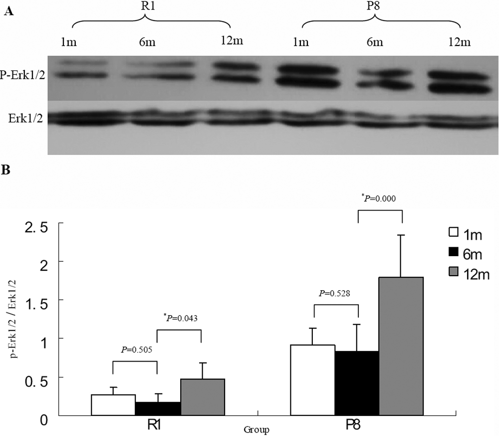
 Figure 9 of
Xiao, Mol Vis 2009; 15:747-761.
Figure 9 of
Xiao, Mol Vis 2009; 15:747-761.