Figure 7. Immunohistochemical staining of p-ERK 1/2, p16INK4a, p19ARF, p21WAF1/CIP1, and p53 in corneal endothelial cells. The arrows indicate the positive staining in the endothelial nuclei at various ages
of each experimental group A: p-ERK 1/2, SAM R1 strain at one month; B: p16INK4a, SAM P8 strain at one month; C: p19ARF, SAM P8 strain at 12 months; D: p21WAF1/CIP1, SAM P8 strain at six months; E: p53, SAM R1 strain at 12 months. F: The negative control incubated with PBS instead of a primary antibody, SAM P8 strain at six months. Magnification: 400X.
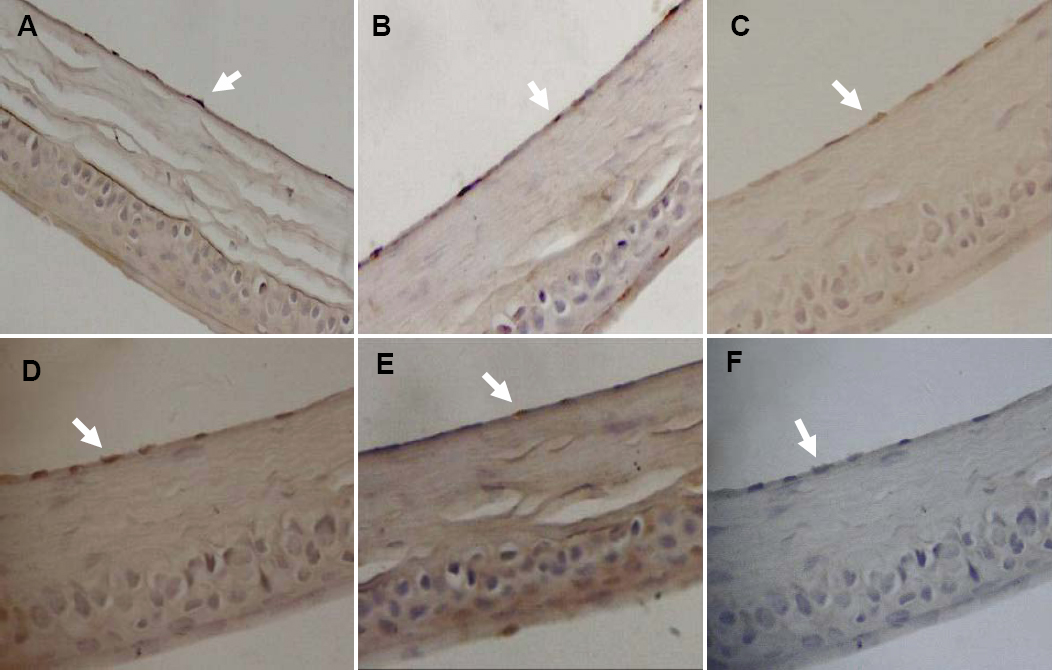
 Figure 7 of
Xiao, Mol Vis 2009; 15:747-761.
Figure 7 of
Xiao, Mol Vis 2009; 15:747-761.