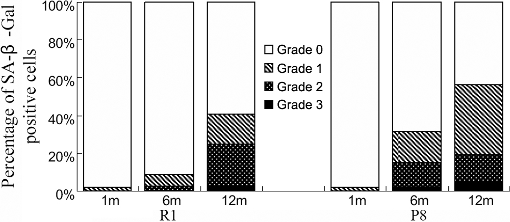
Figure 6. Average percentage of SA-β-Gal-positive cells at each grade in various experimental groups. The classification standard of
SA-β-Gal activity is according to Tatsuya et al. [
49]. This graph shows that few to no positive SA-β-Gal staining was observed in the endothelium in corneas from younger mice.
In corneas from older mice, the percentage of CECs staining positive for SA-β-Gal was higher in the P8 strain than in the
R1 strain.Statistical analysis is shown in
Table 6.
 Figure 6 of
Xiao, Mol Vis 2009; 15:747-761.
Figure 6 of
Xiao, Mol Vis 2009; 15:747-761.