Figure 4. Analysis of scanning electron microscope imaging. Analysis of one-month-old specimens from the SAM R1 and the SAM P8 experimental
groups (A and B, respectively) displayed the morphology of endothelial cells, which was hexagonal and uniform, with numerous microvilli located
on the surface. Analysis of six-month-old specimens from the SAM R1 and the SAM P8 experimental groups (C and D, respectively) showed that the endothelial cells became larger and pantomorphic. Analysis of 12-month-old specimens from
the SAM R1 and the SAM P8 experimental groups (E and F, respectively) showed more polygonal cells, and these cells also became larger. Scale bars: 10 μm.
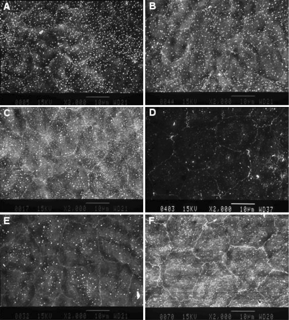
 Figure 4 of
Xiao, Mol Vis 2009; 15:747-761.
Figure 4 of
Xiao, Mol Vis 2009; 15:747-761.