Figure 1. Corneal endothelium of the SAM
strains stained with alizarin red and trypan blue for detection of
the cell morphology, density and viability. A: The endothelium
of a one-month-old specimen from the SAM R1 strain is shown. Most of
the cells are regular hexagon. Magnification: 400X. B: The
endothelium of a six-month-old specimen from the SAM P8 strain shows a
significant number of polygonal cells. Magnification: 400X. C:
The endothelium of a 12-month-old specimen from the SAM P8 strain is
displayed. The arrows indicate the hyperplasia of the tunica vasculosa
on the endothelial surface layer. The covering of the tunica vasculosa
causes most of the cells displayed to look unclear. The blue lines
(black arrow) indicate the hyperplastic blood vessel. Magnification:
100X.
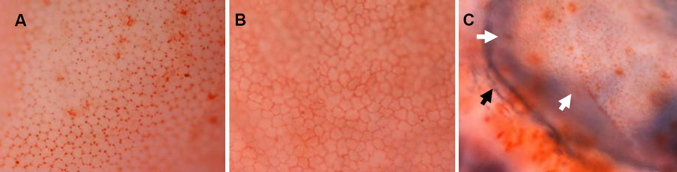
 Figure 1 of Xiao, Mol Vis 2009; 15:747-761.