Figure 4. PKC immunoreactivity in the
smooth muscle layer of the retinal arteries. Representative examples
showing phosphorylated PKCα, PKCβ1, and PKCβ2 immunoreactivity in the
retinal arteries following ischemia and 20 h of reperfusion. Double
staining with smooth muscle actin, a smooth muscle marker, showed
colocalization with phosphorylated PKC in the smooth muscle layer
(arrows). Note that the colocalization was most apparent for
phosphorylated PKCα.
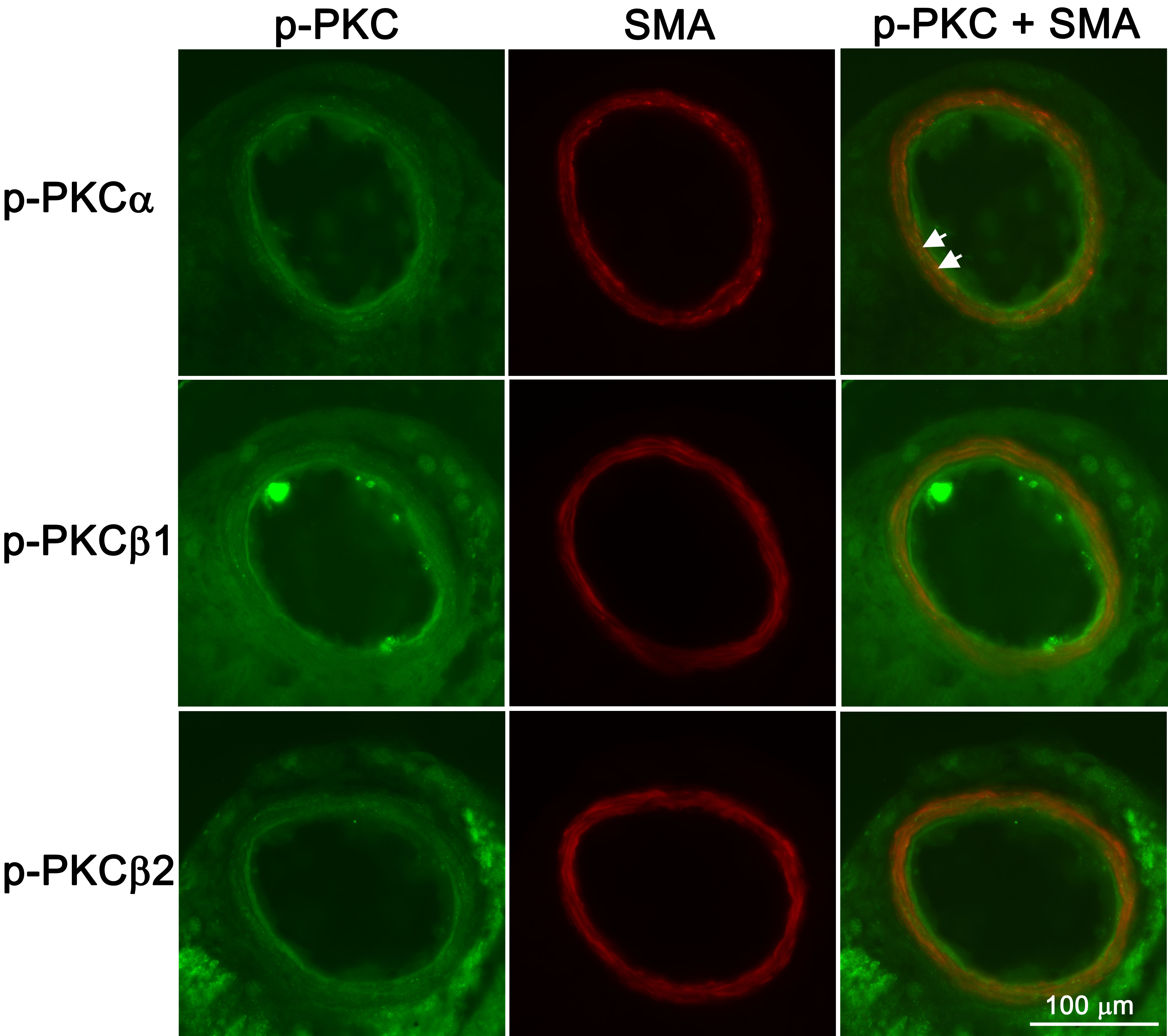
 Figure 4 of Gesslein, Mol Vis 2009; 15:737-746.