Figure 3. PKC immunoreactivity in the
retinal arteries and neuroretina. Representative examples showing
phosphorylated PKCα, PKCβ1, and PKCβ2 immunoreactivity in the retinal
arteries and retina following ischemia and 20 h of reperfusion. A:
Double staining with CD31 (also called PECAM-1), an endothelial cell
marker, showed co-localization of phosphorylated PKC in the endothelium
(arrows). Weak phosphorylated PKC staining could also be seen in the
smooth muscle layer. B: The lower levels of PKCα, PKCβ1, and
PKCβ2 observed in the neuroretina after ischemia–reperfusion, according
to western blot, were reflected in the immunofluorescence staining
results, showing less staining for phosphorylated PKCα and PKCβ2 in the
ischemia–reperfusion eyes compared to sham-operated eyes. Furthermore,
the phosphorylated PKCβ1 staining showed fewer labeled bipolar cells
bodies in the eyes subject to ischemia-reperfusion compared to
sham-operated eyes (see insert in the p-PKCβ1 picture). Similar results
were seen in all pigs studied. Abbreviations: outer nuclear layer
(ONL), outer plexiform layer (OPL), inner nuclear layer (INL), inner
plexiform layer (IPL), ganglion cell layer (GCL), and nerve fiber layer
(NFL).
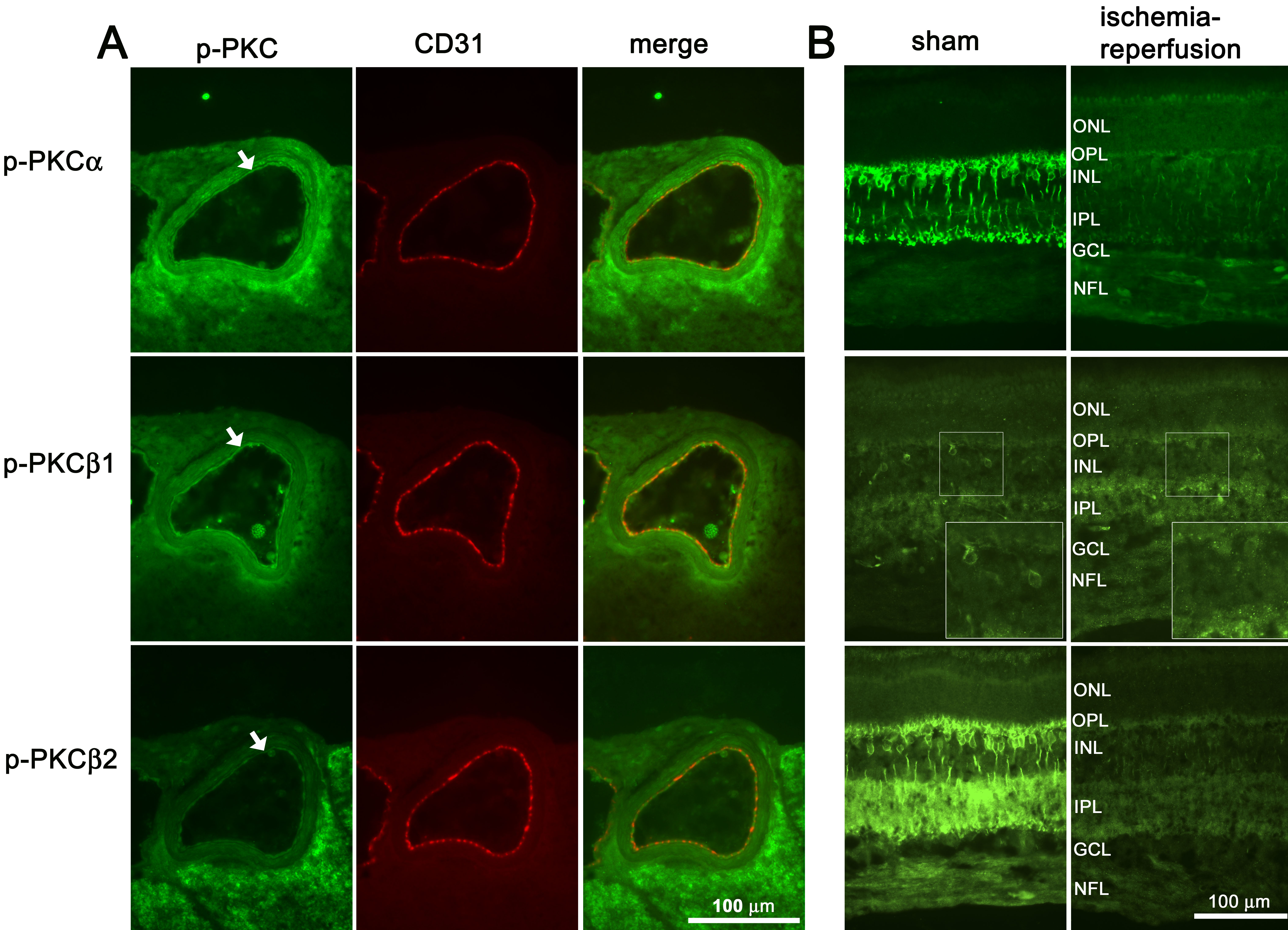
 Figure 3 of Gesslein, Mol Vis 2009; 15:737-746.