Figure 5. NMDA induces CREB phosphorylation in vivo. Photomicrographs show retinal sections collected from postnatal rats 1 h, 2 h,
and 1 day after intravitreal injection of BrdU-saline (Control) or BrdU-NMDA (NMDA). The retinal sections were labeled with
antibodies against P-CREB or BrdU and counterstained with DAPI. Abbreviations: ganglion cell layer (GCL); inner plexiform
layer (IPL); inner nuclear layer (INL); outer plexiform layer (OPL); outer nuclear layer (ONL). Arrow indicates P-CREB immunopositive
cells. Asterisk (*) denotes P-CREB-BrdU coimmunolabeled cells. Pound sign (#) marks BrdU immunopositive cells. Calibration
bar denotes 50 μm in middle left panel.
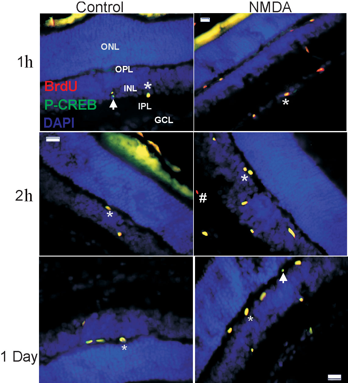
 Figure 5 of
Ramírez, Mol Vis 2009; 15:713-721.
Figure 5 of
Ramírez, Mol Vis 2009; 15:713-721.