Figure 4. Transmission electron microscopy result of paraffin section of the proband’s cornea stained with Hale's colloidal iron. A: The picture shows that the collagen fibers in the corneal stroma are irregular, enlarged, and intensive (1,500X). B: The picture shows hyperplastic, active fibroblasts and intracytoplasmic vacuoles in the cornea (5,000X).
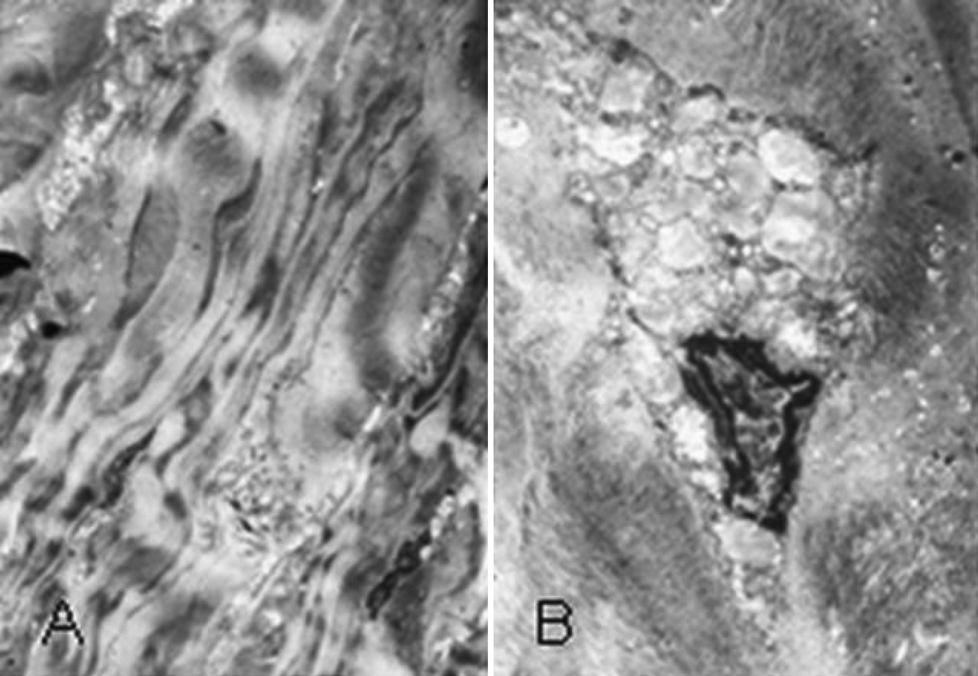
 Figure 4 of
Dang, Mol Vis 2009; 15:700-705.
Figure 4 of
Dang, Mol Vis 2009; 15:700-705.