Figure 3. Paraffin section of the proband’s cornea stained with Hale’s colloidal iron. Blue plaque deposits were found in the corneal
stroma and under the endothelial layer. These deposits presented a significantly positive reaction to colloidal iron staining.
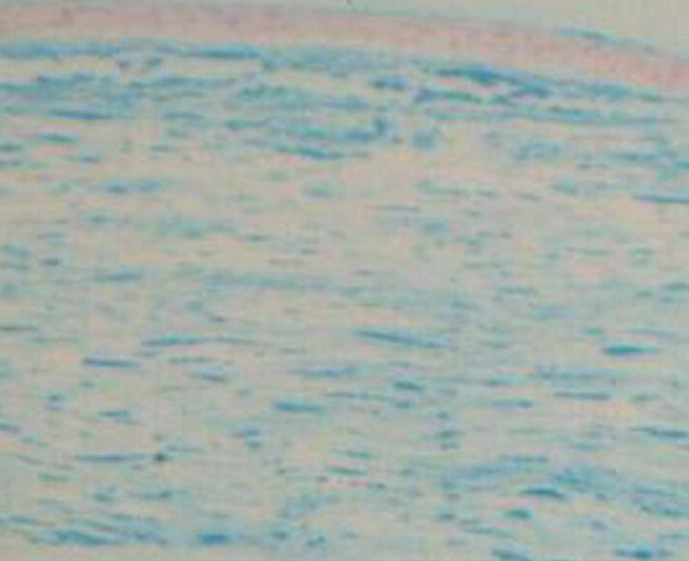
 Figure 3 of
Dang, Mol Vis 2009; 15:700-705.
Figure 3 of
Dang, Mol Vis 2009; 15:700-705.