Figure 5. Paraffin-embedded histopathology
of AND-34−/− mouse eyes. Paraffin tissue sections of
four-month-old AND-34−/− eyes are compared with a
four-month-old AND-34+/+ eye. Panel B shows a
fragment of anterior chamber lens cortex (short arrow) in front of the
iris (long arrow). Panel C shows a fragment of lens cortex
(short arrow) behind the iris (long arrow). Panel D shows that
the fragment of lens cortex lacks both epithelial cells and a lens
capsule. The lens capsule is visible in the lens itself on the left
side of this panel. In panels A and B, the loss of lens
fiber material from the lens is due to artifactual shearing during the
cutting of the paraffin section.
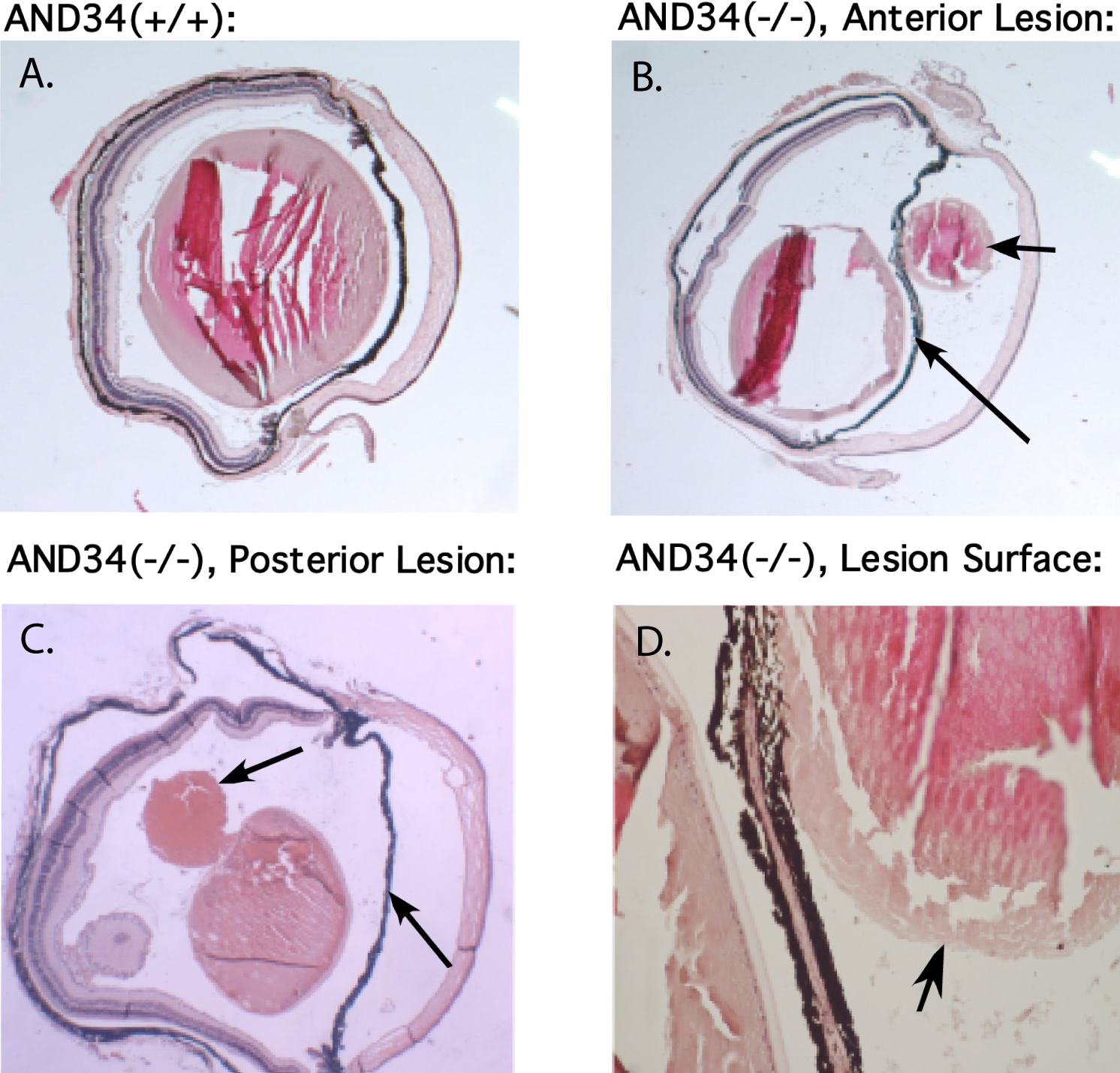
 Figure 5 of Near, Mol Vis 2009; 15:685-699.