Figure 3. AND-34 expression in mouse
tissues. A: Western blot analysis of wild type mouse tissues is
shown. AND-34 was immunoprecipitated directly from lysates of mouse
organs with polyclonal anti-AND-34 antibody whereas p130CAS was
detected directly from whole cell lysates with a monoclonal
anti-p130CAS antibody that is also able to detect HEF1. B: In
situ hybridization for AND-34 transcripts from lymph nodes and
spleens of AND-34+/+ is displayed. The tissues were probed
with sense or antisense hybridization probes as shown in dark-field
microscopy. With dark-field illumination, the positive hybridization
signal of the silver grains in the autoradiographic emulsion appears as
white dots over a black background. C: In situ hybridization
for AND-34 transcripts in the lungs of AND-34+/+
mice is shown in bright-field microscopy. With bright-field
illumination, the positive hybridization signal appears as black dots.
Hybridization was detected in a subset of alveolar cells with the
morphology of lung fibroblasts. Arrows indicate a strong hybridization
signal.
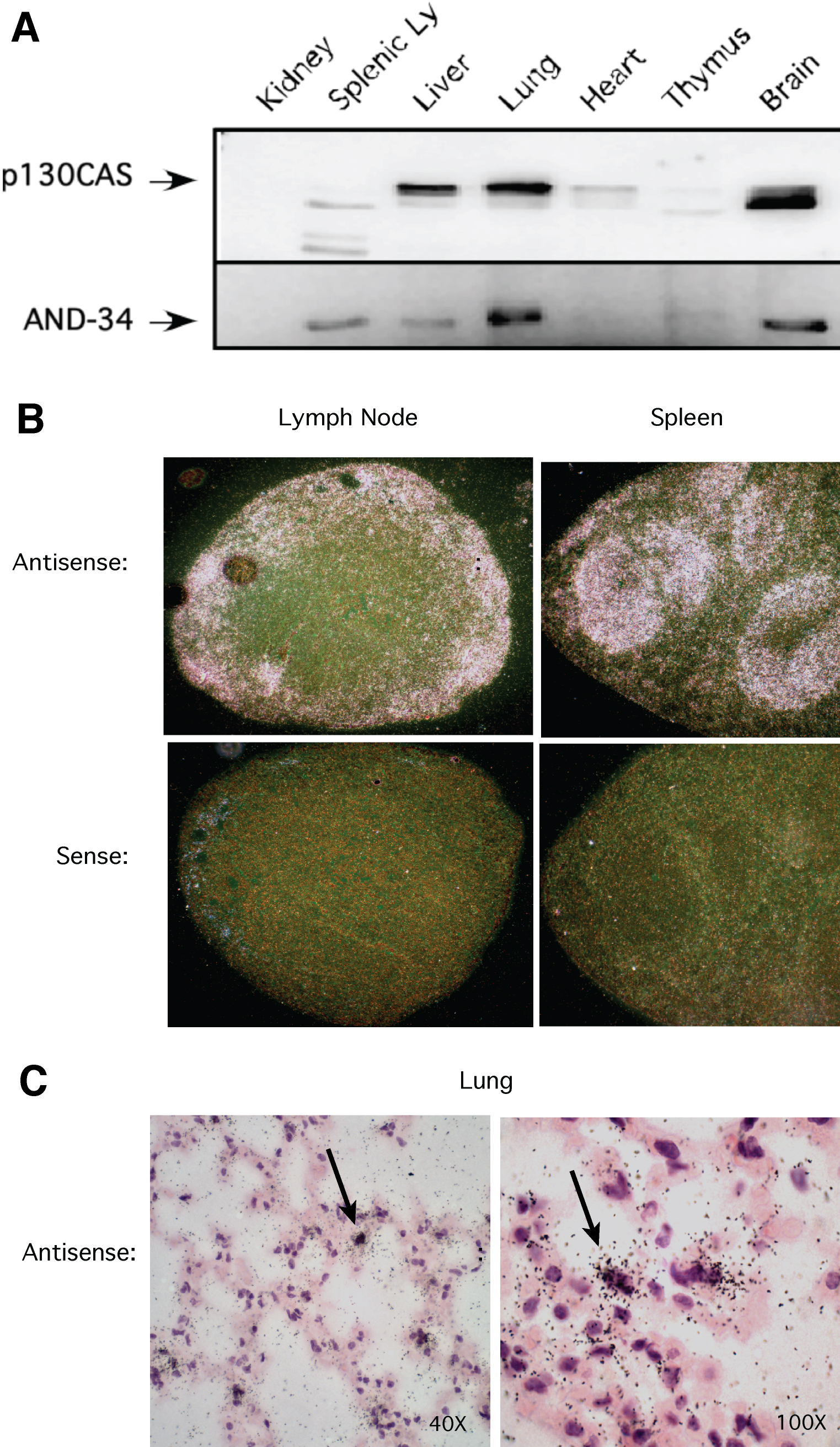
 Figure 3 of Near, Mol Vis 2009; 15:685-699.