Figure 4. Histopathological features of
affected cornea from the right eye of the proband carrying the
homozygous TGFBI R124H mutation at the age of 43. The visual
acuity of the proband before penetrating keratoplasty was 20 cm/CF. A:
Light microscopy of excised corneal tissue stained with congo red
revealed variably sized and irregularly shaped deposits of amyloid
material, predominantly in the anterior and middle stroma (arrows). B:
The deposits stained with Masson’s trichrome showed brightly red
accumulation of hyaline under the Bowman's membrane (arrows). The
epithelium showed focal areas of dehiscence from Bowman's membrane.
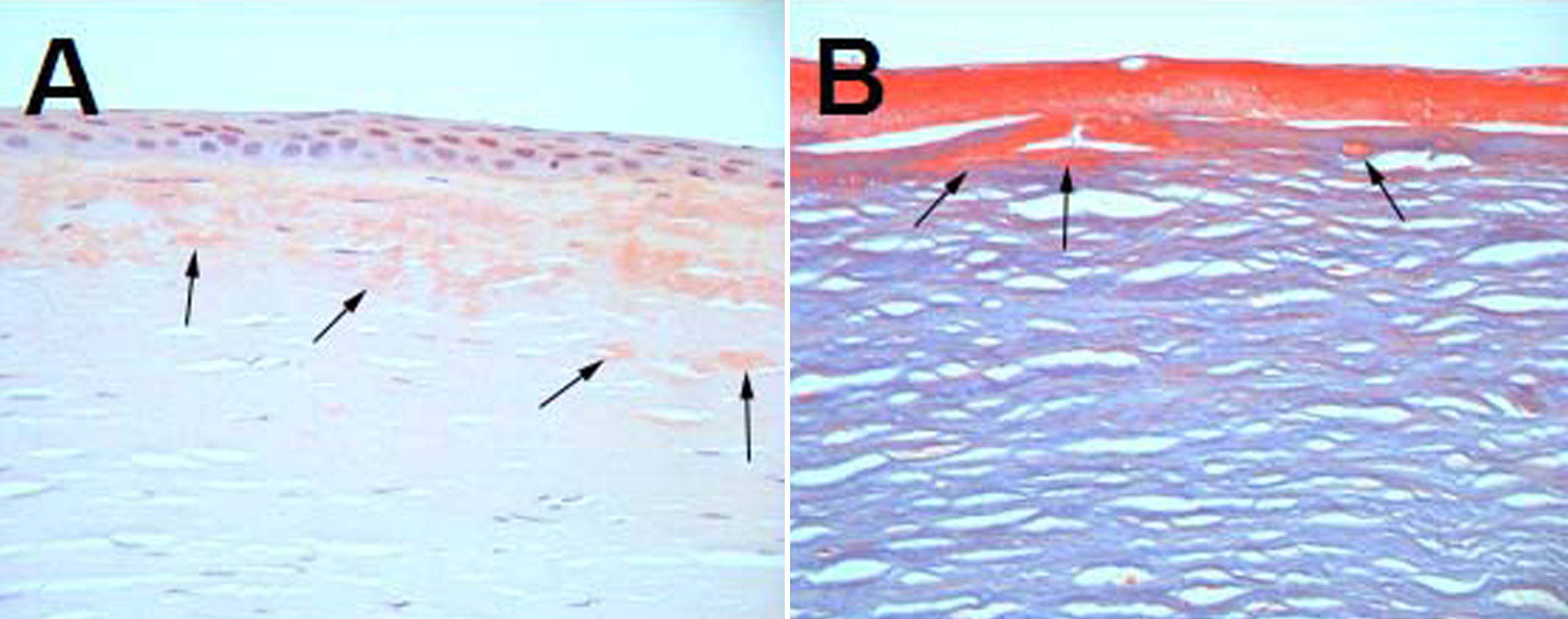
 Figure 4 of Cao, Mol Vis 2009; 15:70-75.