Figure 2. Corneal phenotype of the
affected family members with ACD due to an R124H mutation in TGFBI.
A: Preoperative photograph of the right eye of proband revealed
grayish spot-like confluent opacities presented in the anterior stroma
and covering almost the entire cornea. B: Corneal examination
of the left eye of I1 (father of the proband) showed several distinct
granular deposits in the superficial stroma of the central cornea
(arrows). C and D: Multiple granular deposits and a few
confluent opacities were observed in the anterior stroma of central
cornea in both right eyes of III2 and III5, respectively.
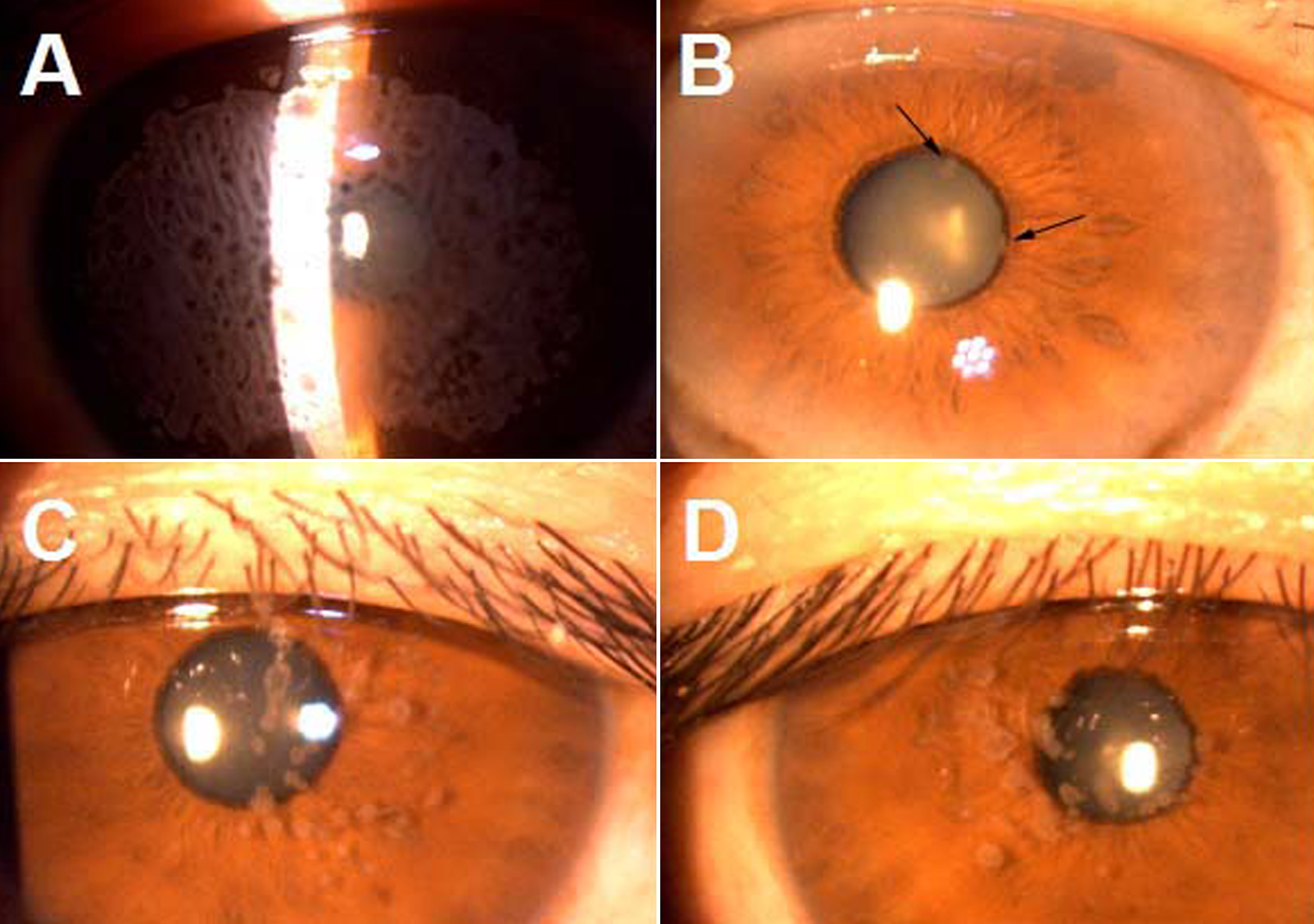
 Figure 2 of Cao, Mol Vis 2009; 15:70-75.