Figure 5. Cellular reactions after photocoagulation in young and old mice. Representative phase contrast images (A, C) and immunohistochemistry (B, D) at month 7 after photocoagulation in 10–12-week-old mice (group III; A, B) and 1-year-old mice (group IV) (C, D). A, C: Phase contrast images show a thickened choroid with pseudocystic cavities (black arrows). B, D: Immunofluorescence analysis was performed in 10–12-week-old mice (B) and 1-year-old mice (D). Choroidal neovascularization reaction is more prominent with aging (yellow arrows). Immunohistochemistry demonstrates that
some of the pseudocystic cavities are vessels (white arrows). Scale bar (A-D) represents 100 µm. Abbreviations: choroid (ch), ganglion cell layer (gcl), inner nuclear layer (inl), outer nuclear layer
(onl), rod outer segments (ros), retinal pigment epithelium (rpe), sclera (scl).
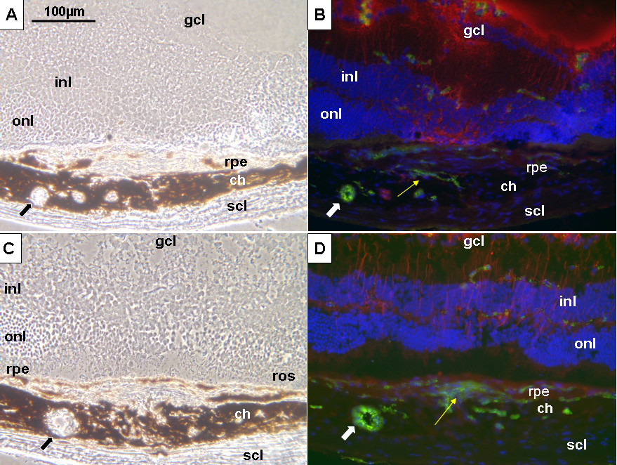
 Figure 5 of
Dot, Mol Vis 2009; 15:670-684.
Figure 5 of
Dot, Mol Vis 2009; 15:670-684.