Figure 4. Representative phase contrast
image and immunohistochemistry four months after photocoagulation in
1-year-old mice (group IV). A: Phase contrast image
demonstrates a capsular and pigmented cell reaction that closes the
initial retinal pigment epithelium (rpe) interruption and surrounds the
choroidal neovascularization (CNV) reaction. Overlying the lesion,
photoreceptor outer segments are markedly affected. B:
Immunodetection of von Willebrand factor (green) reflects the CNV
presence at least four months after photocoagulation limited by the
surrounding new RPE. The new vessels do not infiltrate the retina.
Astrocytes and activated Müller cells were detected with specific
antibodies for glial fibrillary acidic protein (GFAP, red). Nuclei were
counterstained with 4’,6-Diamidino-2-Phenyl-Indole (DAPI, blue). Scale
bar in A represents 100 µm in both panels. Abbreviations:
choroid (ch), ganglion cell layer (gcl), inner nuclear layer (inl),
outer nuclear layer (onl), rod outer segments (ros), retinal pigment
epithelium (rpe), sclera (scl).
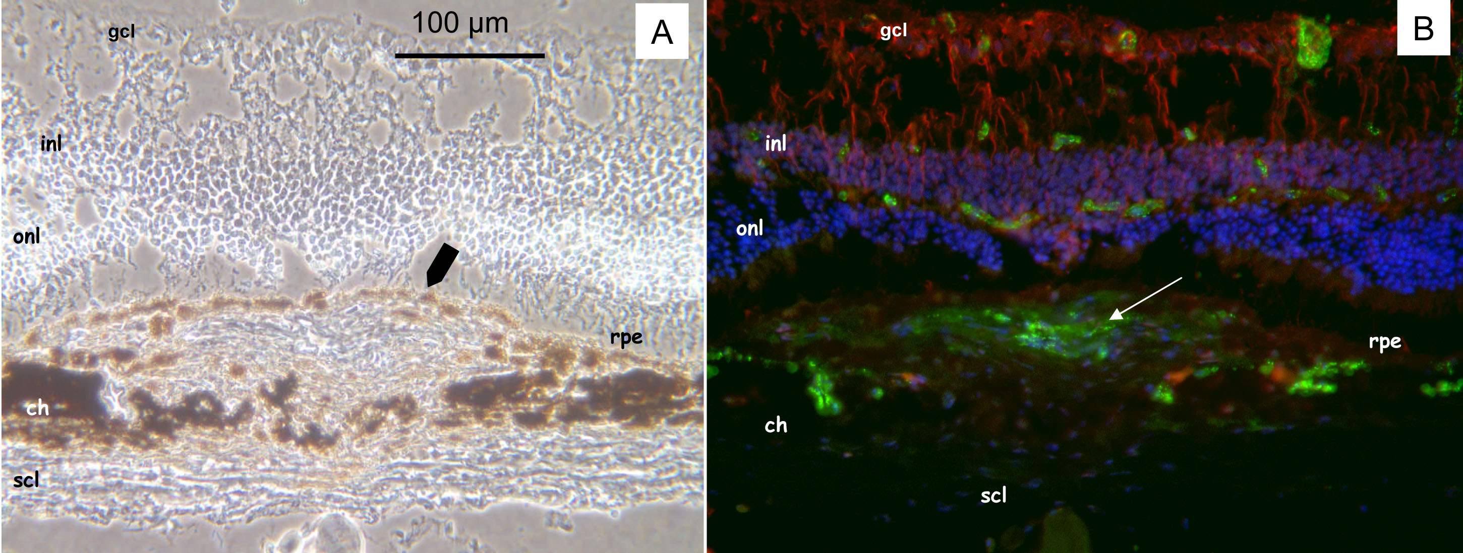
 Figure 4 of Dot, Mol Vis 2009; 15:670-684.