Figure 3. Size of RPE interruption after
photocoagulation. Size of RPE interruption was evaluated for mice from
the four groups (group I: 4 weeks, group II: 6 weeks, group III: 10–12
weeks, group IV: 12 months), from day 7 to month 4 after
photocoagulation. A: The size of retinal pigment epithelium
(RPE) interruption is maximal on day 7 after the laser burn in the four
groups and decreases thereafter. It is larger in the 10–12-week-old
mice (in green) at each time point. From month 4 a repair of the RPE
layer is evident in 1-year-old mice; no interruption is recorded. B:
The mean RPE interruption during the first two weeks after
photocoagulation was as follows: 136.9 µm±4.4 in group I, 174.1 µm±6.6
in group II, 219.1 µm±12.2 in group III, and 112.1 µm±11.4 in group IV.
The differences observed in the RPE interruption between mice from
groups I, II III and mice from group IV are significant (p<0,0001)
according to the t-test.
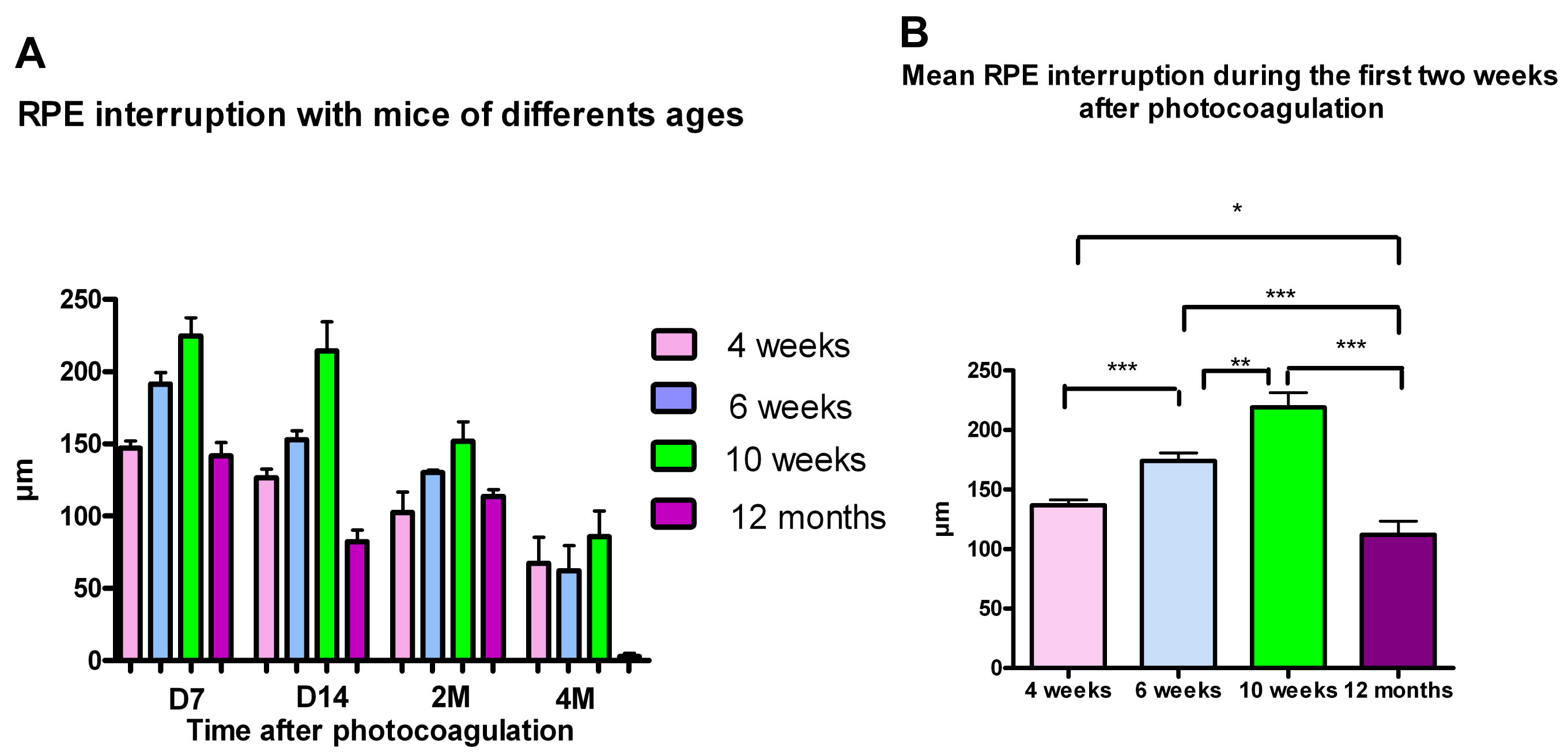
 Figure 3 of Dot, Mol Vis 2009; 15:670-684.