Figure 10. MCP-1 expression after
photocoagulation in RPE-choroid complex. A: The amount of
monocyte chemotactic protein-1 (MCP-1) transcripts was compared
with 18S transcripts (y-axis) expressed in the same samples.
These data are from four different eyes. Values in histogram are
means±SEM B,C: Representative samples of PCR fragments
(individual ratio of MCP-1:18S closest to the corresponding mean) for
10–12-week-old mice (B) and 1-year-old mice (C) were
analyzed by 3% agarose gel electrophoresis and visualized by ethidium
bromide staining under ultraviolet light. Prior to laser treatment
MCP-1 mRNA expression was very low. After photocoagulation, it is
strongly expressed in the retinal pigment epithelium-choroid complex;
its level of expression is higher and observed for a longer period of
time in old mice.
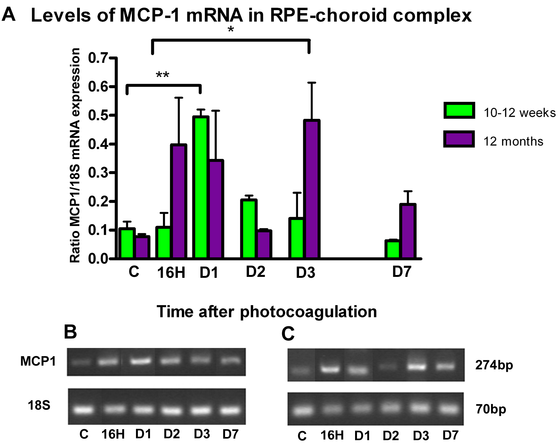
 Figure 10 of Dot, Mol Vis 2009; 15:670-684.