Figure 1. Cellular reactions after
photocoagulation in 10–12-week-old mice (group III). Representative
phase contrast images and immunohistochemistry of sections from eyes at
day 7 (A, C), and month 6 (B, D) following
photocoagulation. A: Phase contrast image demonstrates a
localized disruption of retinal pigmented epithelium (rpe) and choroid
(ch) in the center of the laser beam (arrows). B: Phase
contrast image demonstrates the formation of a mound of poorly defined
cells surrounded by lightly pigmented retinal pigmented epithelium
(rpe) structures (arrow). C: Immunohistofluorescence analysis
was performed with specific antibodies for von Willebrand (green) and
glial fibrillary acidic protein (GFAP, red). Nuclei were counterstained
with 4’,6-Diamidino-2-Phenyl-Indole (DAPI, blue). This section also
illustrates a marked activation of GFAP-positive cells (astrocytes and
Müller cells) in the center of the laser beam and von Willebrand
reaction in the choroidal site (thin arrows). D: Six months
after photocoagulation, a localized von Willebrand reaction is still
evident at the level of the choroid (thin arrows). Abbreviations:
ganglion cell layer (gcl), inner nuclear layer (inl), inner plexiform
layer (ipl), outer nuclear layer (onl), sclera (scl). Scale bar (A)
represents 100 µm for each panel.
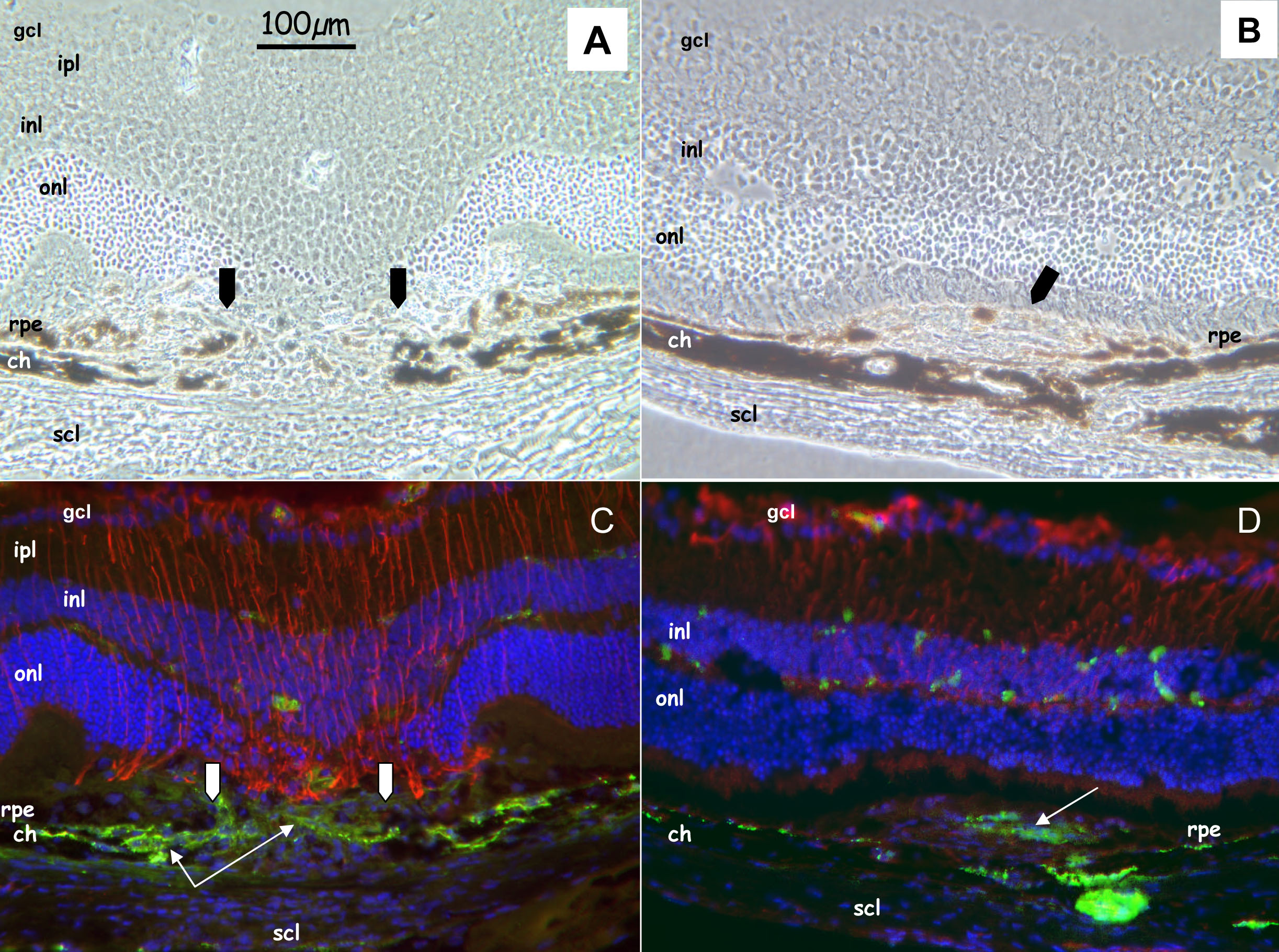
 Figure 1 of Dot, Mol Vis 2009; 15:670-684.