Figure 5. Colocalization of BDNF with GFAP
by double immunofluorescence. A: Schematic drawing shows the
coronal section through the level of the superior colliculus (SC;
bregma −3.40 mm) in mice. The boxed area is the region of the
superficial layer in the contralateral SC. Representative photographs
show brain-derived neurotrophic factor (BDNF; B and C)
and glial fibrillary acid protein (GFAP; D and E)
immunostaining, and BDNF/GFAP (F and G)
double-immunostaining of the contralateral SC at 30 days after
intravitreal N-methyl-D-aspartate (NMDA) injection in mice.
Some BDNF-expressing cells (B and C, green) were
colocalized with GFAP-positive astroglial cells (D and E,
red), as indicated by the yellow color in F (merge of B
and D) and G (merge of C and E). The
scale bars represents 50 µm (B, D, and F) or 10 µm (C,
E, and G).
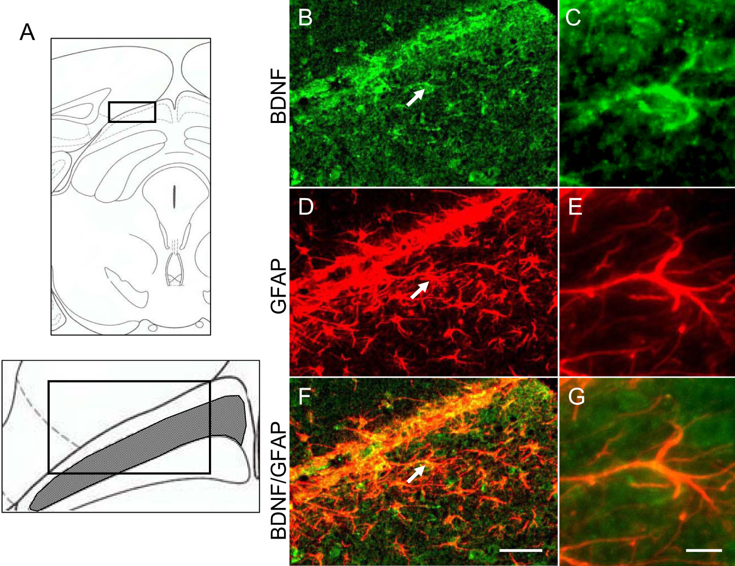
 Figure 5 of Tanaka, Mol Vis 2009; 15:662-669.