Figure 1. The projection pathway from the
retina to the SC. Illustrations show (A) the pathway from the
retina to the superior colliculus (SC), (B) the coronal section
through the level of the SC (bregma−3.40 mm) in mice; boxed areas
contain the SC of the contralateral side and the ipsilateral side and (C)
the superficial layer of the SC.
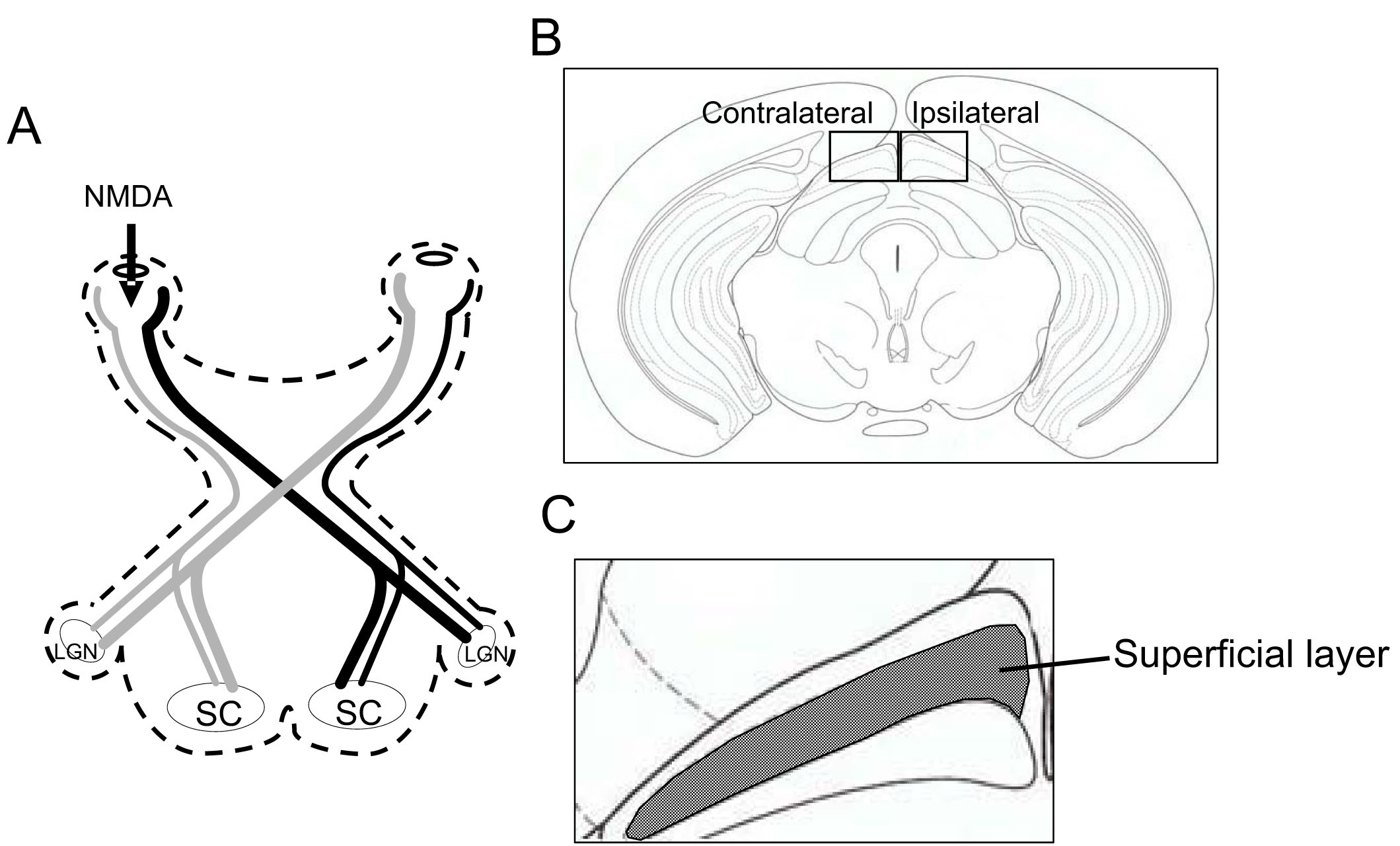
 Figure 1 of Tanaka, Mol Vis 2009; 15:662-669.