Figure 1. Typical retinal phenotype in
affected members A: Left fundus of proband showing temporal
optic nerve pallor, attenuated retinal blood vessels, macular
pigmentary and atrophic changes, and some fine pigment clumping outside
the vascular arcade. B: Mid-transit fluorescein angiogram of
left fundus of younger sister reveals a dark choroid sign with some
retinal pigment epithelium (RPE) transmission defects in a perifoveal
distribution. C and D: Right and left fundus
photographs of younger sister at age 12 years show mottling of RPE in
macular region but no flagrant pigmentary changes; there is slight
temporal pallor of the optic nerve heads, especially in left eye.
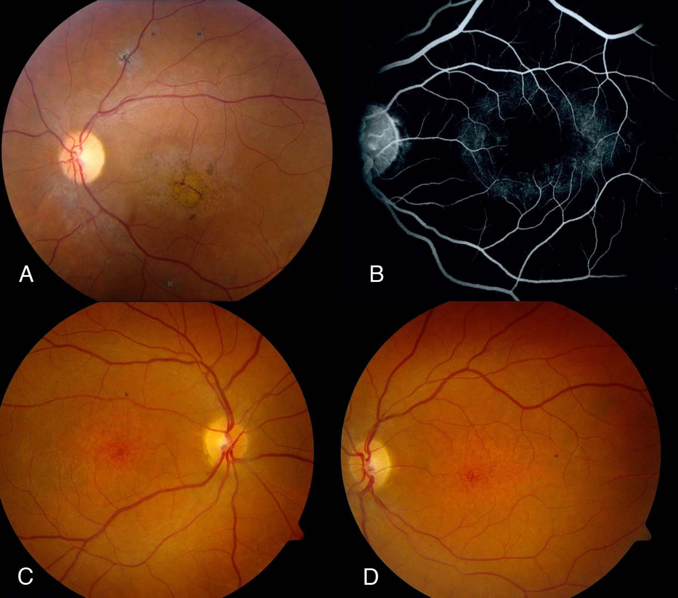
 Figure 1 of Xi, Mol Vis 2009; 15:638-645.