Figure 6 of
Engler, Mol Vis 2009; 15:629-637.
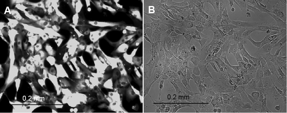
Figure 6.
Human corneal endothelial cells transfected with Amaxa using pmaxGFP on day 3 after FACS. Fluorescence microscopy (
A
), exhibits an essentially pure population of transfected cells as compared with corresponding bright-field image (
B
).
 Figure 6 of
Engler, Mol Vis 2009; 15:629-637.
Figure 6 of
Engler, Mol Vis 2009; 15:629-637.