Figure 5 of
Engler, Mol Vis 2009; 15:629-637.
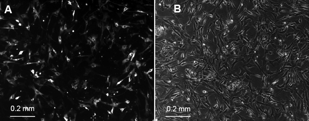
Figure 5.
Human corneal endothelial cells on day 5 after transfection with Amaxa. Fluorescence microscopy (
A
) shows a transfection rate of ~30% compared with corresponding bright-field image (
B
).
 Figure 5 of
Engler, Mol Vis 2009; 15:629-637.
Figure 5 of
Engler, Mol Vis 2009; 15:629-637.