Figure 2 of
Engler, Mol Vis 2009; 15:629-637.
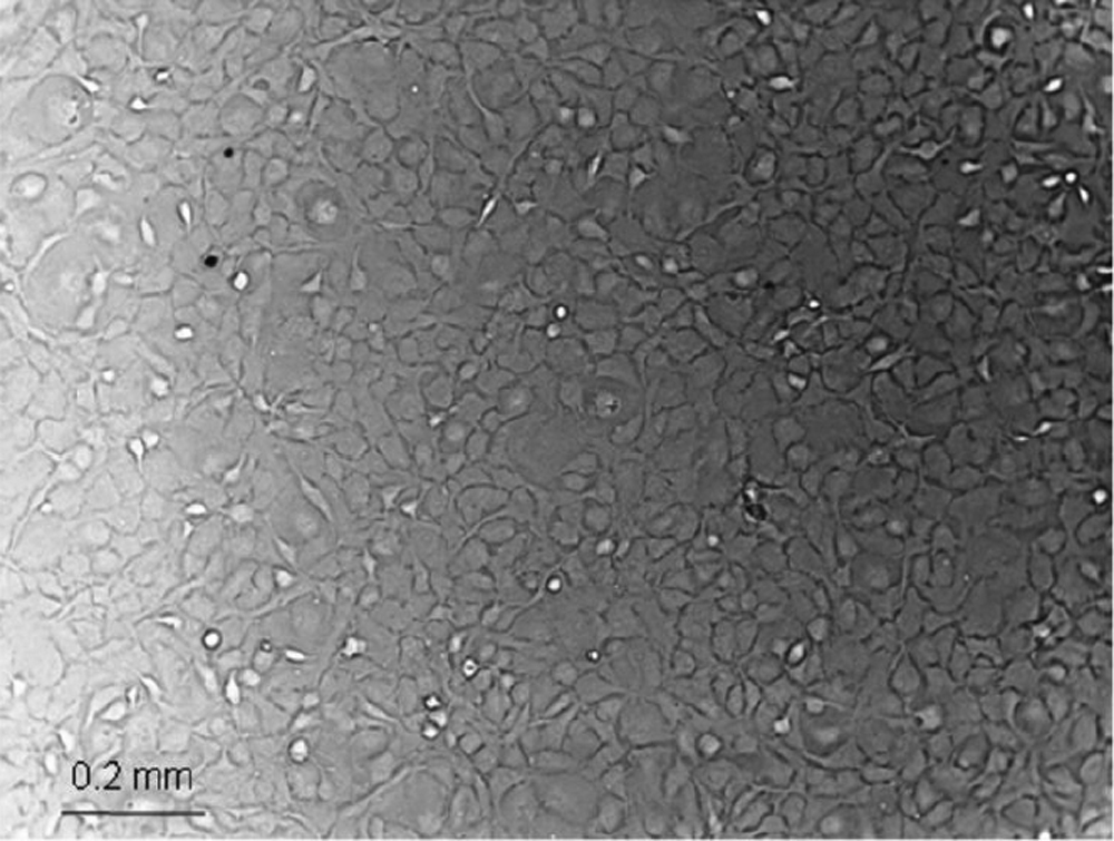
Figure 2.
Human corneal endothelial cells after 10 days of culturing before first passage. Cells exhibited a polygonal shape similar as in vivo.
 Figure 2 of
Engler, Mol Vis 2009; 15:629-637. Figure 2 of
Engler, Mol Vis 2009; 15:629-637.
Figure 2 of
Engler, Mol Vis 2009; 15:629-637. Figure 2 of
Engler, Mol Vis 2009; 15:629-637.