Figure 5. CD163+ cells
displayed different morphologies at different layers in the iris 48 h
after LPS injection. This figure shows different stromal layers from
the epithelial to endothelial (from A to F) in the same
field. Dendritiform cells are located in the stroma adjacent to the
epithelial layer (A) while round-pleiomorphic cells are adjacent
to the endothelial layer (F). The cells that arrows and
arrowheads point to represent the same cells, respectively. Original
magnification: A–F 400X.
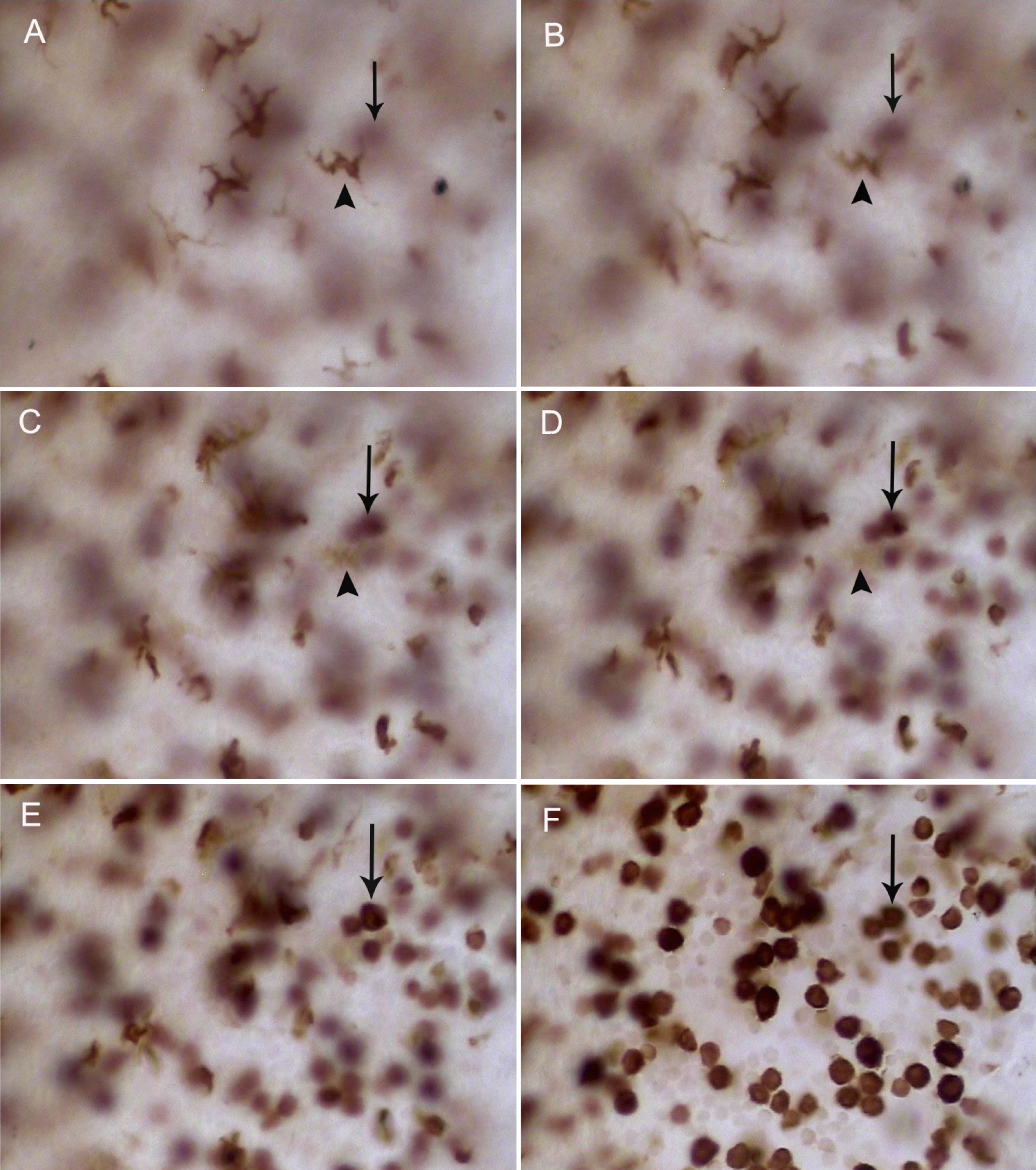
 Figure 5 of Chen, Mol Vis 2009; 15:619-628.