Figure 1. Clinical manifestation of EIU in
Wistar rats. A: The image shows the eye 24 h after the LPS
injection. Note the fibrinous pupillary membrane (arrow). B:
The image shows the eye 48 h after the LPS injection. The fibrinous
pupillary membrane has been absorbed (arrow).
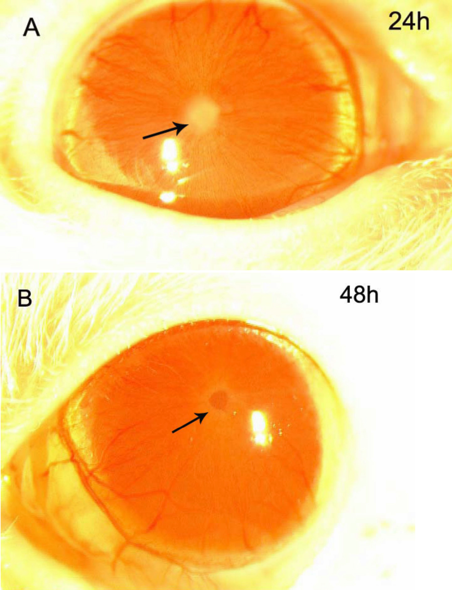
 Figure 1 of Chen, Mol Vis 2009; 15:619-628.