Figure 4. Patient's encephalic MRI. MRI of the optic nerve showed severe bilateral atrophy of the optic nerve from retina to the lateral
geniculate nucleus, seen on horizontal MRI section (A), including atrophy of the optic chiasma, seen on coronal MRI section (B).
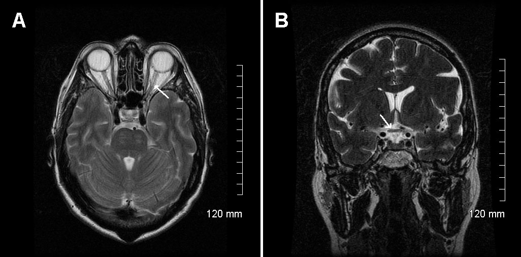
 Figure 4 of
Nochez, Mol Vis 2009; 15:598-608.
Figure 4 of
Nochez, Mol Vis 2009; 15:598-608.