Figure 2 of
Nochez, Mol Vis 2009; 15:598-608.
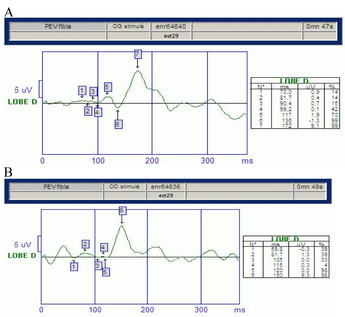
Figure 2.
Patient 's visual evoked potentials. We observe low-amplitude potentials and increased latency in visual evoked potentials in left eye (
A
) and in right eye (
B
). It indicates defective conduction of the optic nerves.
 Figure 2 of
Nochez, Mol Vis 2009; 15:598-608.
Figure 2 of
Nochez, Mol Vis 2009; 15:598-608.