Figure 1 of
Nochez, Mol Vis 2009; 15:598-608.
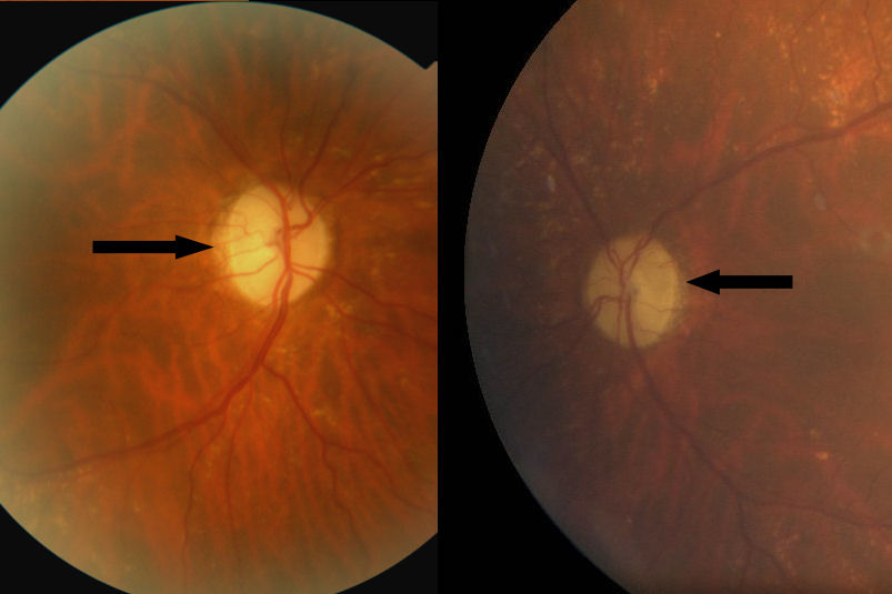
Figure 1.
Fundus examination. The first image represents the patient's right eye fundus examination and we observe optic disk atrophy (arrow). The second image represents left eye fundus examination, we also observe optic disk atrophy.
 Figure 1 of
Nochez, Mol Vis 2009; 15:598-608.
Figure 1 of
Nochez, Mol Vis 2009; 15:598-608.