Figure 5. Dendritic cell distribution
across the ocular surface of Aire-deficient mice. A:
Immunofluorescence study of CD11c+ cells (red) demonstrates
dendritic antigen presenting cells (APCs) in the basal epithelium (open
arrowhead) and sub-epithelial stroma (arrow) of the conjunctiva (Cj),
limbus (Lm), and central cornea (Co). Although CD11c+ cells
are found in both Aire+/− and Aire−/−
mice, significantly more are seen in Aire-deficient mice. CD11c+
cells are present throughout the whole layer of the cornea including
the posterior stroma (solid arrow head) in Aire−/−
while they are largely absent in the posterior cornea in Aire+/−.
The dotted line represents the epithelial basement membrane in the
limbus, and the blue nuclear counterstaining (DAPI) is used for
orientation. Bar, 100 μm. B: Immunohistochemical study of MHC
class II surface antigen (I-Ad) reveals an intense
infiltration of activated dendritic APCs in the epithelium (open arrow
head) and stroma (arrow) of the conjunctiva (Cj), limbus (Lm), and
central cornea (Co) of Aire−/− mice. By comparison,
I-Ad+ cells are rarely apparent in the conjunctiva and
limbus and are completely absent in the central cornea of Aire+/−
mice. The asterisk denotes goblet cells. Bar, 100 μm. C:
Quantification of CD11c+ and I-Ad+
cells is shown over time. Data are shown as mean±SD. An asterisk
indicates that p<0.05, CD11c+Aire+/−
versus CD11c+Aire−/−. The symbol, †,
indicates that p<0.05, I-Ad+Aire+/−
versus I-Ad+Aire−/−.
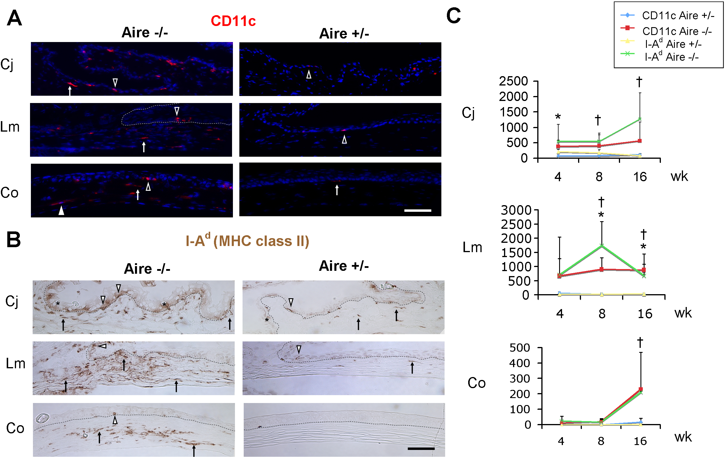
 Figure 5 of Chen, Mol Vis 2009; 15:563-576.