Figure 1. Infiltration and destruction of
the lacrimal gland and ocular epithelium in Aire-deficient
mice. A: Immunohistochemical analysis of CD4+ and CD8+
T cells was negative in Aire+/− while Aire−/−
mice showed intensive multifocal aggregates of both cell types. Of
note, characteristic interlobular connective tissue that exists in the
normal lacrimal gland (asterisks) disappears in the Aire−/−
lacrimal gland following lymphocytic infiltration. Bar, 100 μm. B:
Time course of lissamine green staining in the corneas of Aire+/−
and Aire−/− mice demonstrates a compromised
epithelial integrity caused by Aire deficiency. Mild to severe
keratoconjunctivitis sicca (KCS) shown as punctate to confluent green
staining were observed in Aire−/− mice whereas
corneas of Aire+/− largely remained unstained. C:
Progression of lissamine scores is shown on a chart. Data are shown as
mean±SD. The asterisk in this panel indicates that p<0.05, Aire+/−
versus Aire−/− at each time point.
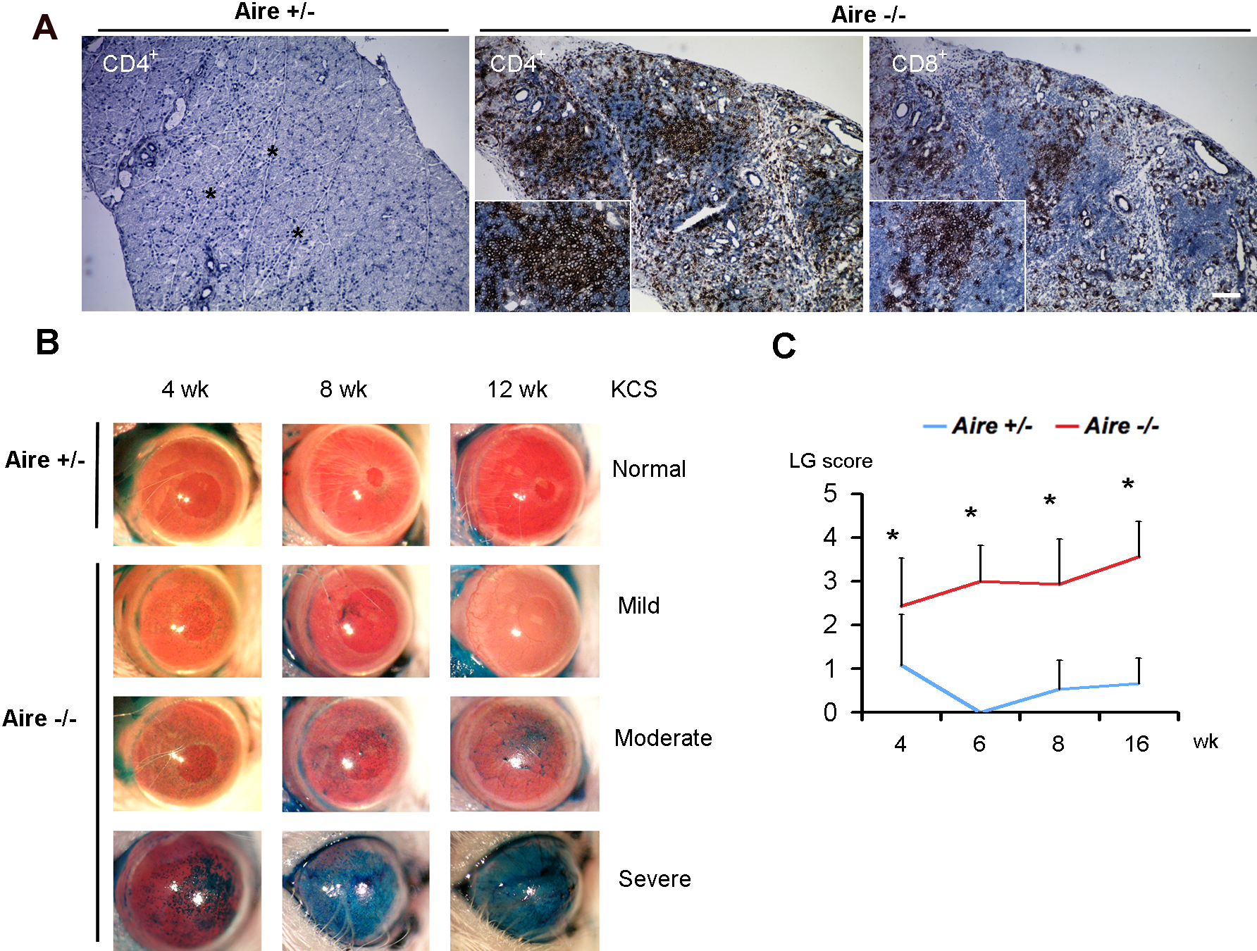
 Figure 1 of Chen, Mol Vis 2009; 15:563-576.