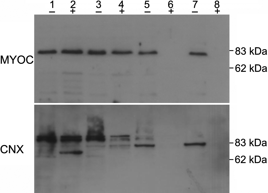
Figure 5. Determination of the topology of myocilin in the endoplasmic reticulum. The microsomal membrane was prepared from HTM cells
transduced with Ad-myocilin-GFP. The membrane fractions were untreated (-) or treated with 1% Triton X-100 (+) followed by
the addition of increasing concentrations of protease K (10 μg/ml in lane 1 and 2; 20 μg/ml in lane 3 and 4; 50 μg in lane
5 and 6; 100 μg lane 7 and 8). After digestion, the membrane was probed with anti-myocilin antibody (upper panel), stripped,
and re-probed with anti-calnexin antibody (lower panel).

 Figure 5 of
Sohn, Mol Vis 2009; 15:545-556.
  Figure 5 of
Sohn, Mol Vis 2009; 15:545-556.