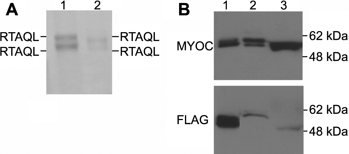
Figure 2. Characterization of the signal sequence cleavage of myocilin.
A: HTM cells were transduced with Ad-myocilin-FLAG and myocilin in cells (lane 1), and culture media (lane 2) were separated
by SDS–PAGE, transferred to the membrane, and loaded into a protein sequencer. All myocilin bands yielded the same results
with respect to their NH
2-terminal amino acid sequences.
B: HTM cells were transduced with Ad-FLAG-myocilin and myocilin in the cells (lane 2), and culture media (lane 3) was detected
by western blot analysis using anti-myocilin antibody (upper panel) or anti-FLAG antibody (lower panel). Myocilin-FLAG (
Figure 1A, lane 2) was included as a control (lane 1).
 Figure 2 of
Sohn, Mol Vis 2009; 15:545-556.
Figure 2 of
Sohn, Mol Vis 2009; 15:545-556.