Figure 5. Characterization of cells in retinas from 12-week-old miR-182 KO mice. A-C: Ganglion and amacrine cells were labeled using anti-Pax6 antibodies in wild-type (+/+), heterozygous (+/−) and homozygous
(−/−) KO mice. D-F: Photoreceptor cells were labeled using anti-recoverin antibodies. G-I: Müller cells were labeled using anti-GS antibodies. J-L: Rod bipolar cells were labeled using anti-PKCα antibodies. Primary antibody labeling is depicted in red. Lower panels show
merged labeling patterns with DAPI (green).
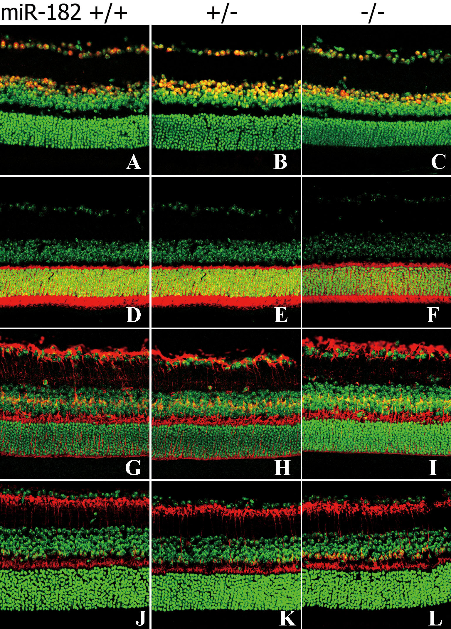
 Figure 5 of
Jin, Mol Vis 2009; 15:523-533.
Figure 5 of
Jin, Mol Vis 2009; 15:523-533.