Figure 6. Distribution of FAK in mouse
cornea after keratectomy. Keratectomy was performed in the center of
corneas of wild type mice and healed for 3 weeks. The excised corneas
were subjected to whole mount electroimmunostaining as described in
Methods. CLSM revealed that anti-FAK Alexa 555 conjugate specifically
labeled the cytoplasmic membrane of stromal cells three weeks after
keratectomy. FAK is shown in red. Scale bar: 10 μm
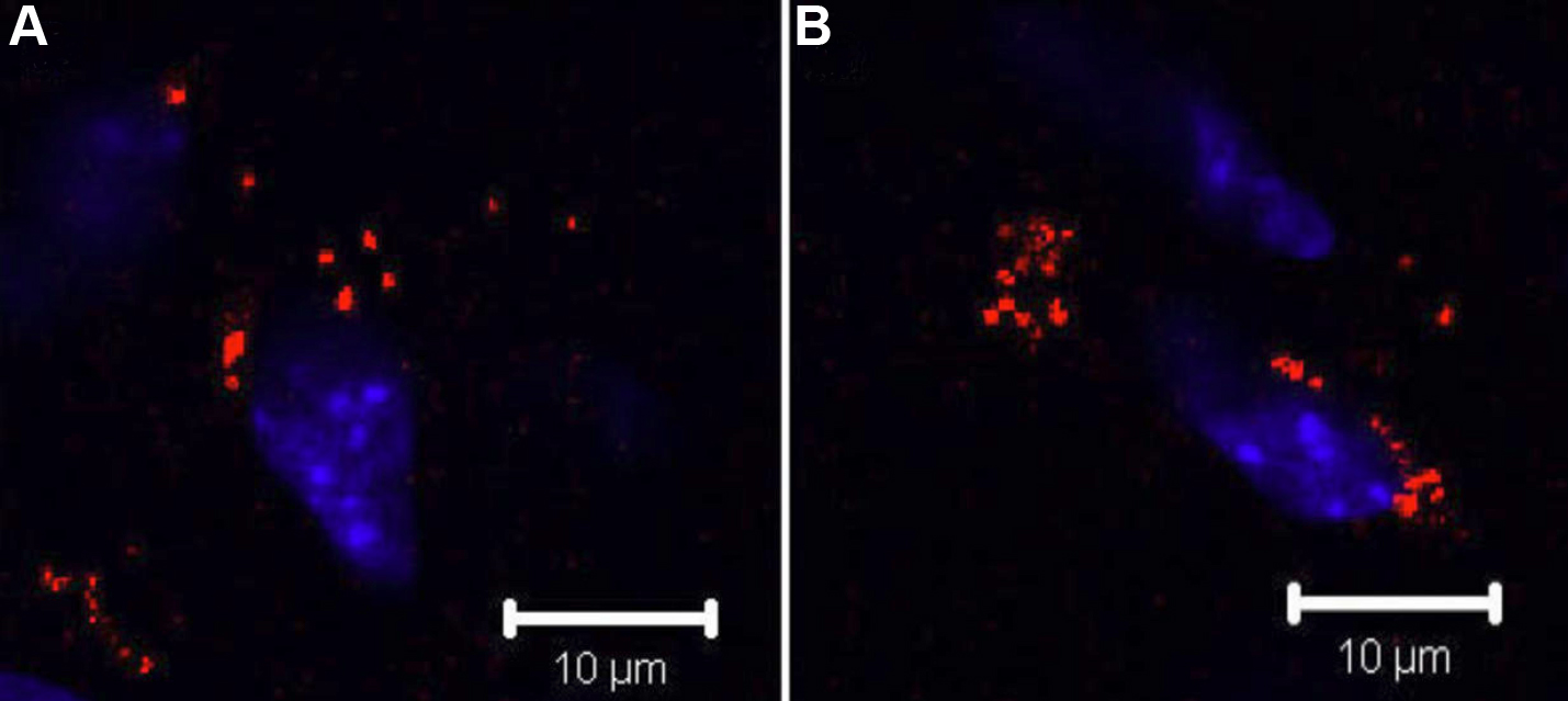
 Figure 6 of Liu, Mol Vis 2009; 15:505-517.