Figure 5. Distribution of F-actin in the mouse corneal cells after the conventional whole mount staining of phalloidin. In the superficial
epithelium, F-actin (red) is homogeneously distributed in the whole cell body (A). In the corneal basal and wing cell layer, the microfilament networks are mostly gathered in the cytoplasmic circumference
(B). In the corneal stroma, phalloidin labeling displays large and flat dendritic cell body of keratocyte (C). In the corneal endothelium, the distribution of F-actin gathered in the circumferential cytoplasm similar to that of the
basal epithelial cell layer (D). Scale bar: 10 μm.
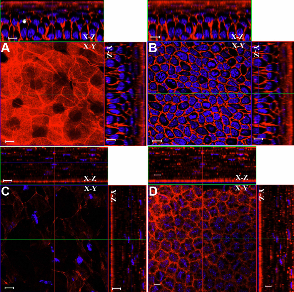
 Figure 5 of
Liu, Mol Vis 2009; 15:505-517.
Figure 5 of
Liu, Mol Vis 2009; 15:505-517.