Figure 4. Distribution of F-actin in the
mouse corneal cells and formation of filopodia and lamellipodia during
healing of epithelium debridement. In the superficial epithelium,
F-actin (red) is homogeneously distributed within whole cell body (A).
However, in the corneal basal and wing cell layer, the microfilament
networks are mostly gathered in the cytoplasmic circumference (B).
In the corneal stroma, phalloidin labeling displays large and flat
dendritic cell body of keratocyte (C,D). In the corneal
endothelium, the distribution of F-actin gathered in the
circumferential cytoplasm similar to that of the basal epithelial cell
layer (E).The squamous epithelial cells at the leading edge
altered the polygonal cell shape into spindle-shape and scallop-shape
that oriented onto the substratum with scallop-shaped lamellipodia and
filopodia. Polymorphonuclear neutrophils invading the stroma following
corneal epithelium debridement were readily visible (F). Scale
bar: 10 μm.
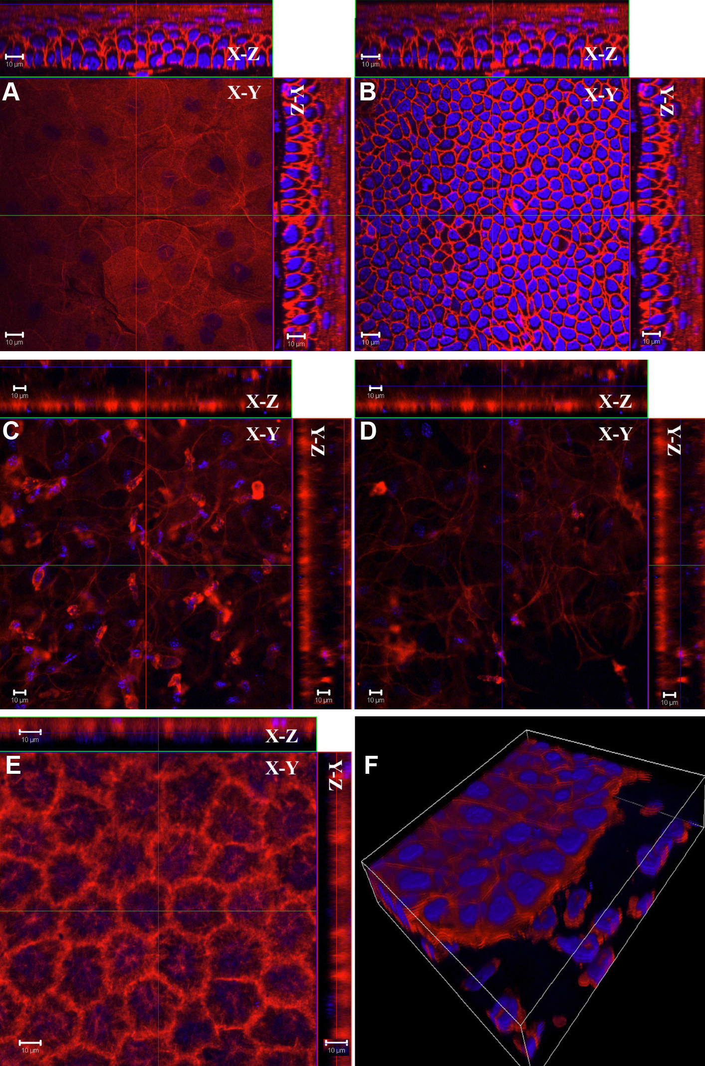
 Figure 4 of Liu, Mol Vis 2009; 15:505-517.