Figure 2. Whole mount
electro-immunofluorescent staining with anti-keratocan. Wild type mouse
corneal stroma was labeled by goat anti-keratocan Alexa555 conjugate
(red) and nuclei were stained with DAPI (blue) as described in Methods.
The red fluorescence is only detected in corneal stroma (A). No
red fluorescent signals are found in the keratocan knockout mouse
cornea by goat anti-keratocan Alexa555 conjugate (B). The wild
type mouse corneal stroma is not labeled by non-immune goat IgG
Alexa555 conjugate (C). Conventional whole mount fluorescent
immunostaining with anti-keratocan-Alexa 555 conjugate showed that the
antibody couldn’t penetrated into the deep corneal stroma and exhibited
non-specific binding on the surface of the corneal epithelium and
endothelium (D). Scale bar: 10 μm.
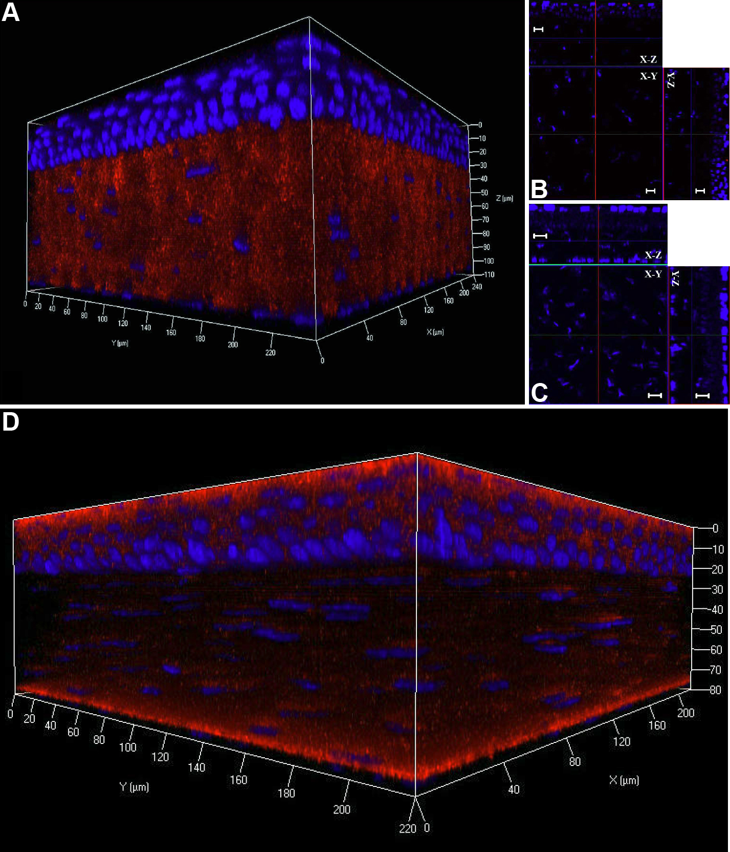
 Figure 2 of Liu, Mol Vis 2009; 15:505-517.