Figure 1. Diagrams of the electrophoresis
apparatus. A: The column is made of plastic tubing, the inner
diameter is about 6 mm, and the length is 15 mm. The column
consisted of three layers. The first layer contains a cornea (or other
tissue) embedded in 1% agarose in 0.05% Triton X-100 in TGB. The second
layer is 0.5% agarose containing IgG fluorescent conjugate in TGB, and
the third layer is 2% agarose in TGB. B: Depending on
predetermined net charges of the reagent in TGB (pH 7.4), the column
was directionally immersed in a submarine gel electrophoresis apparatus
filled with 1X TGB, and then the IgG conjugates were electrophoresed
into the cornea at 4–10 mA.
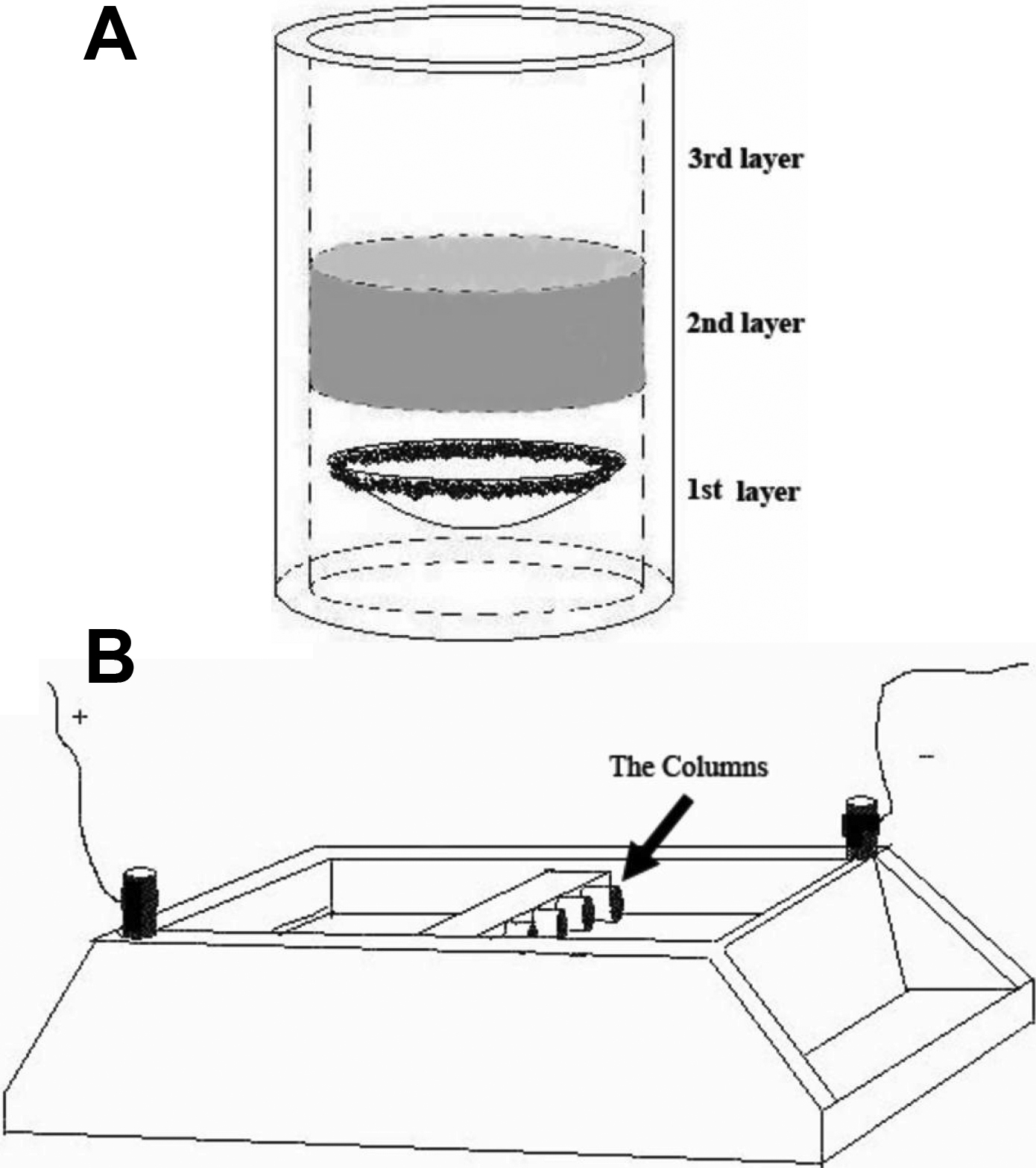
 Figure 1 of Liu, Mol Vis 2009; 15:505-517.