Figure 4. Increased retinal ganglion cell
survival in the RGS2−/− mice. To determine the effect of RGS2 on
retinal ganglion cell survival in the RGS2−/− mice, we used retinal
cryosections derived from 12 to 15-month old male mice and
immunolabeled ganglion cells with Brn-3-specific antibody. The same
specimens were also stained with Hoechst 33258 dye to count the total
number of cells in the ganglion cell region. Panels A and B
show Brn-3-specific immunostaining (red), C and D show
Hoechst staining (blue), and E and F depict the
overlapping of Brn-3 and Hoechst staining. G: Quantitative
changes in the Brn-3-positive retinal ganglion cells between the
RGS2−/− and wild-type mice. H: Quantitative changes in the
total number of cells in the ganglion cell region of the retina in
RGS2−/− and wild-type mice. The RGS2−/− mice show a significant
increase in Brn-3-positive cells in the ganglion cell region of the
central retina as compared to the wild-type mice. The total number of
cells in the ganglion cell region of the central retina based on
Hoechst staining did not show significant difference between two groups
of the mice. Cell number is expressed per unit area, and values are
expressed as mean±SEM (n=4). The asterisk indicates a p<0.05.
Abbreviations: ganglion cell layer (GCL); inner plexiform layer (IPL);
inner nuclear layer 563 (INL); outer nuclear layer (ONL); outer
plexiform layer (OPL). Scale bar represents 100 μm.
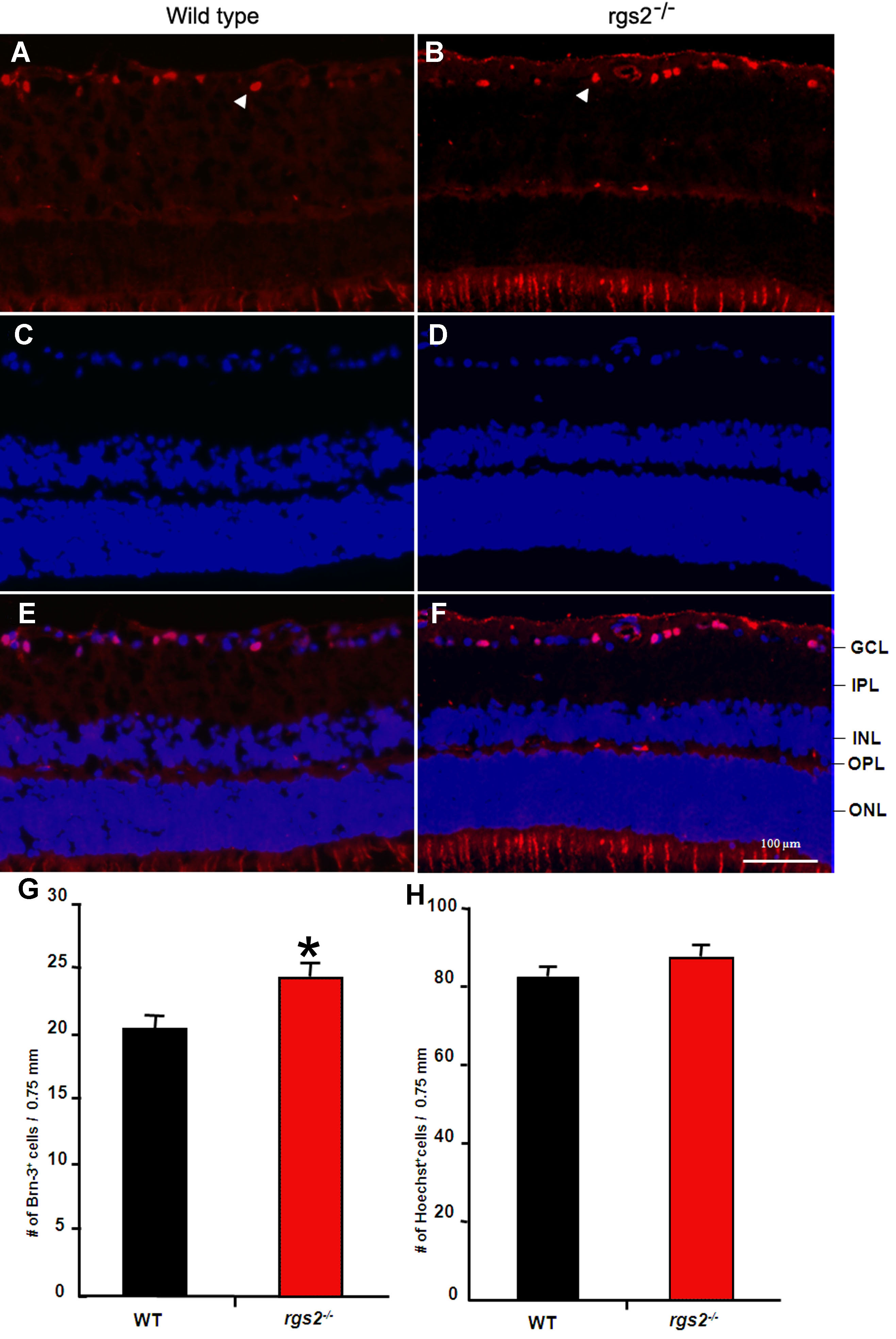
 Figure 4 of Inoue-Mochita, Mol Vis 2009; 15:495-504.