Figure 3. Changes in actin filament
staining at the iridocorneal angle of the RGS2−/− mice.
Cryosections of the eye anterior chamber including the iridocorneal
region derived from the RGS2−/− and wild-type mice were
labeled for filamentous actin with rhodamin-phalloidin, and
fluorescence images were recorded. Actin filament staining (bright red
fluorescence) in the ciliary muscle (CM) and trabecular meshwork (TM)
of the RGS2−/− mice (C and D) was found to be
more intense relative to the wild-type mice (A and B),
with much stronger staining in the ciliary body (CB) as opposed to the
TM. Panels E-H show images of the same specimens stained for
the actin filaments (A-D), but recorded under bright-field
settings. Abbreviations: retina (RT); Schlemm’s canal (SC); sclera
(SCL). Scale bar represents 50 µm.
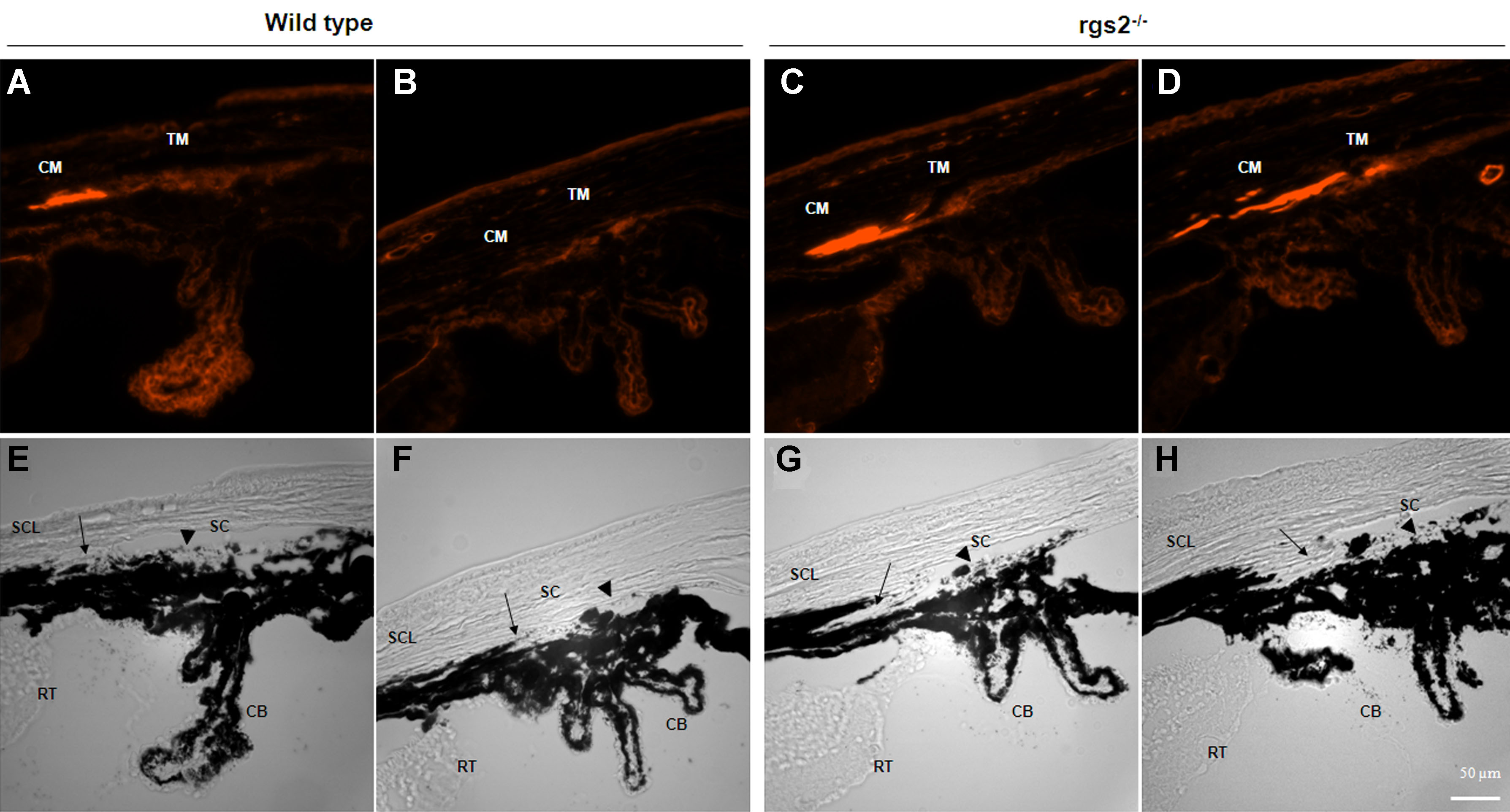
 Figure 3 of Inoue-Mochita, Mol Vis 2009; 15:495-504.