Figure 2. Iridocorneal histological
changes in the RGS2−/− mice. A: Iridocorneal
histological analysis was performed to examine structural differences
in the aqueous humor outflow pathway and ciliary body between the RGS2−/−
and wild-type mice. The structural integrity of the trabecular meshwork
(TM), ciliary body (CB), and ciliary muscle (CM) were found to be
comparable between the two groups of mice. However, the Schlemm’s canal
(SC) area was found to be significantly larger in the RGS2−/−
mice compared to the littermate wild-type mice. B: Quantitative
changes in the SC area between the RGS2−/− and wild-type
mice based on Metamorph pixel analysis. Values are shown as mean±SEM
(n=6). Asterisk represents p<0.05. Abbreviations: sclera (SCL),
retina (RT).
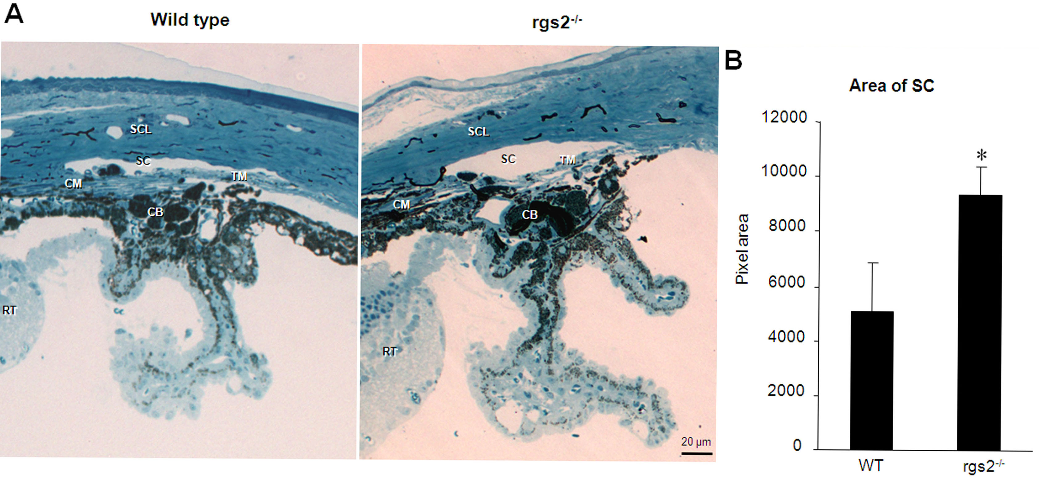
 Figure 2 of Inoue-Mochita, Mol Vis 2009; 15:495-504.