Figure 1. Intraocular pressure changes in
the RGS2−/− mice. Diurnal IOP was measured in both right and
left eyes in conscious animals, using a rebound tonometer. Measurements
were performed five times on the same animals at weekly intervals A-E:
IOP changes in the RGS2−/− mice were compared with those of
wild-type (WT) mice recorded over five different days. F: IOP
changes between the wild-type and RGS2−/− mice, based on the
average of five independent measurements, are shown in panel A-E.
Each error bar represents the mean±SEM (n=8–10 eyes). Asterisk
represents p<0 0.05.
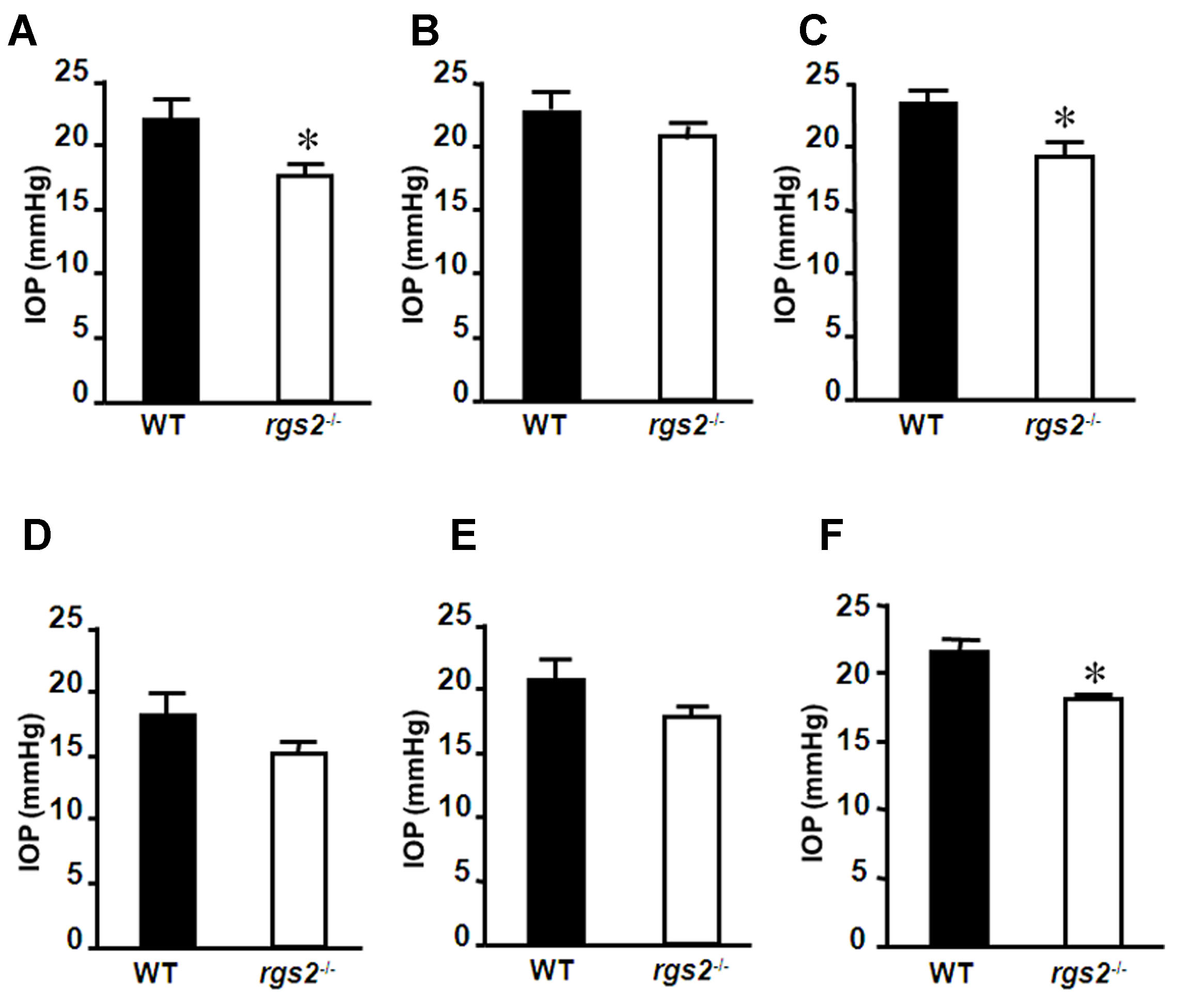
 Figure 1 of Inoue-Mochita, Mol Vis 2009; 15:495-504.