Figure 4. In situ hybridization for PEDF
mRNA (dark reaction product) in fetal macaque retina. Layers of the
retina are indicated by the vertical brackets. Vessel profiles are
indicated in C and D by oblique arrows. A, B:
At Fd 80, higher levels of PEDF expression were detected in the
GCL at the incipient fovea (A) compared with a location near the
optic disc (B). PEDF levels were low in the RPE at Fd
80. Labeled cells in the IPL of A are most likely displaced
ganglion cells which are common in central retina at this age. C,
D: In the Fd 150 fovea (C), relatively high levels of PEDF
mRNA are present in the GCL compared with levels in the GCL near the
optic disc (D). Inset shows increased PEDF expression in
the RPE at Fd 150 compared with Fd 80 (see A and B).
Abbreviations: ganglion cell layer (GCL); inner nuclear layer (INL);
inner plexiform layer (IPL); outer nuclear layer (ONL); outer plexiform
layer (OPL); nerve fiber layer (NFL); retinal pigmented epithelium
(RPE).
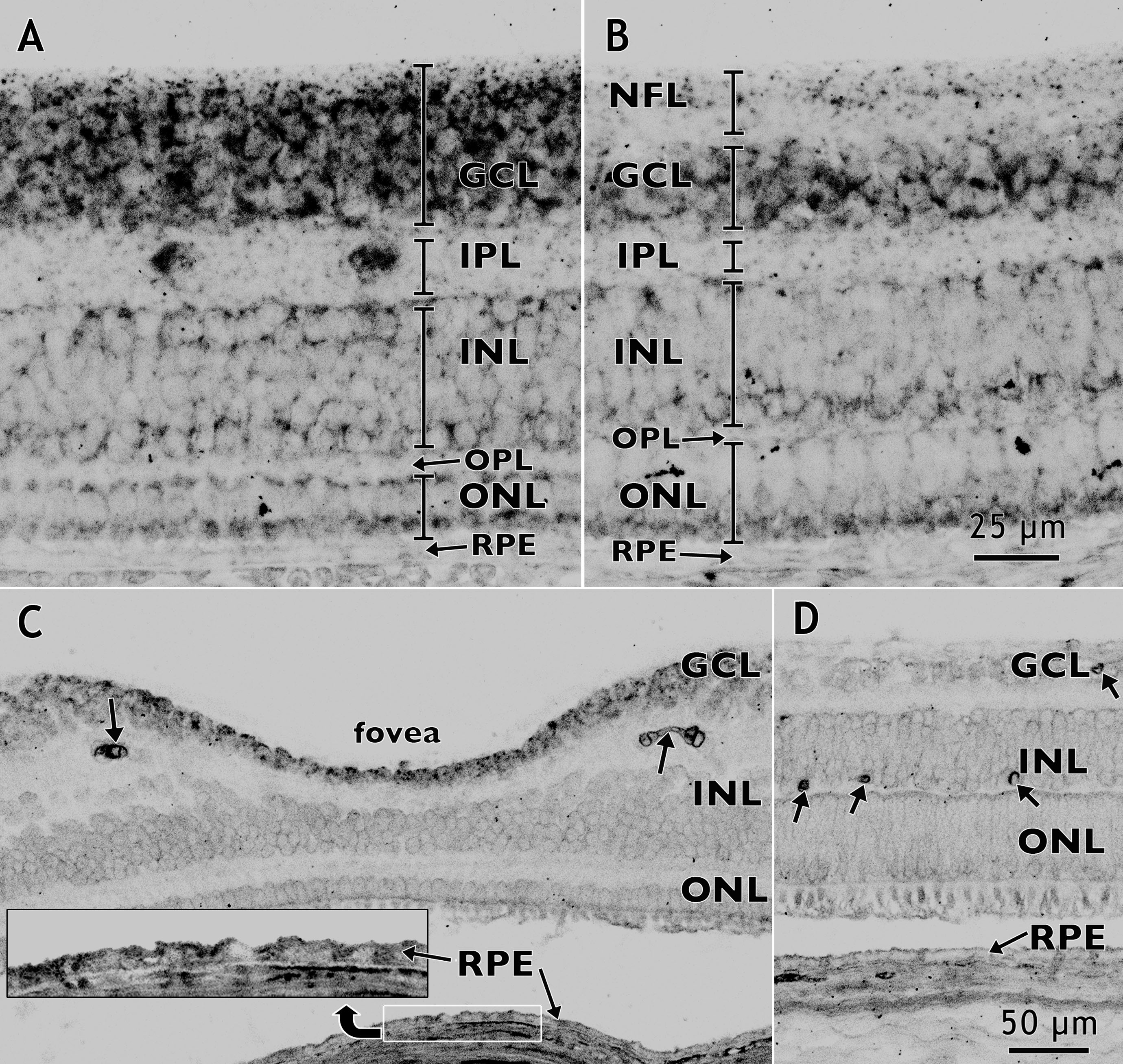
 Figure 4 of Kozulin, Mol Vis 2009; 15:45-59.