Figure 3. In situ hybridization for Eph-A6
mRNA in fetal macaque retina. Small arrows indicate the profiles of
retinal vessels, and the thicker arrows (to the left) mark the vessels
on the margin of the foveal avascular zone (FAZ). Double-headed arrows
show optical densitometry transects (B-D). A:Eph-A6
mRNA expression is shown in a section through the developing fovea in a
retina from a macaque at Fd 115. There are high levels of Eph-A6
expression (dark reaction product) in the inner part of the ganglion
cell layer (GCL) at the developing fovea and lower levels of Eph-A6
expression in the GCL at progressively more peripheral locations,
consistent with both the QRT–PCR and microarray findings. A second
gradient of Eph-A6 mRNA expression across the depth of the GCL
at foveal and parafoveal locations (left and central of A) was
also detected, as shown by optical densitometry profiles at B, C,
and D. B: A transect through the retina within the FAZ
shows peak expression of Eph-A6 mRNA in the inner GCL,
declining to low levels in the outer GCL. C: A weaker gradient
was detected across the GCL about 1.5 mm from the incipient fovea,
with peak levels approximately 70% of the peak value in the fovea. D:Eph-A6
expression was generally lower in the periphery, although slightly
higher levels are present in the GCL compared with the other layers.
Values in C and D are normalized to the peak intensity
in B. Abbreviations: arbitrary units (AU); inner nuclear layer
(INL); outer nuclear layer (ONL).
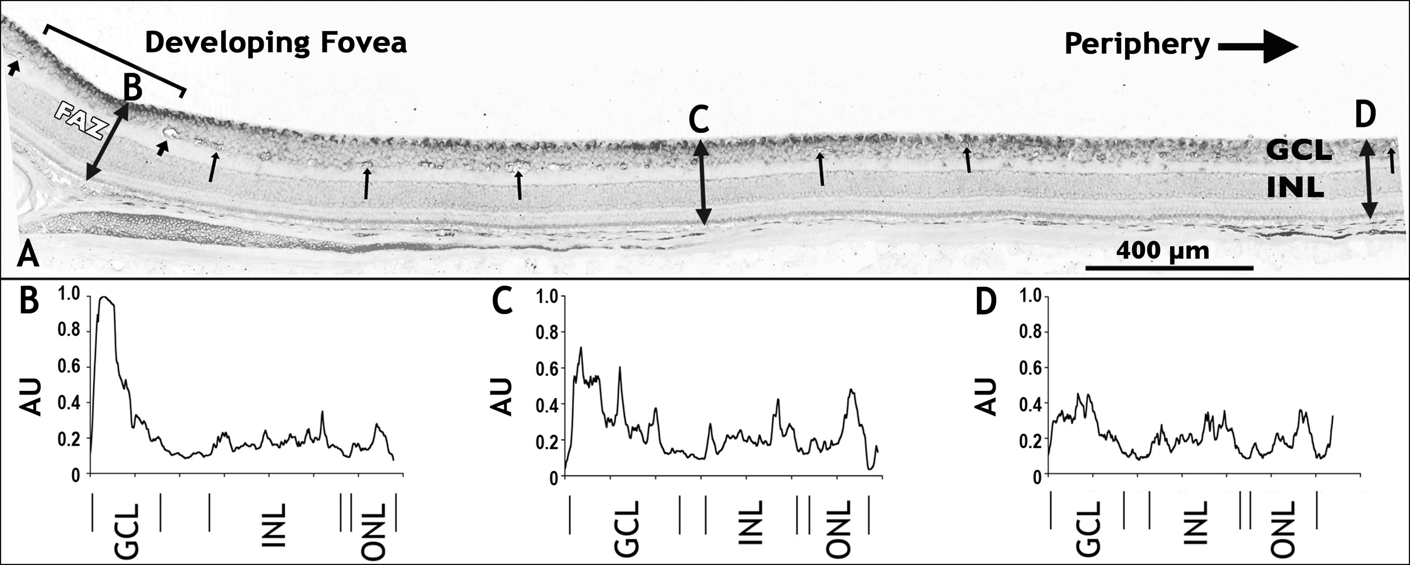
 Figure 3 of Kozulin, Mol Vis 2009; 15:45-59.