Figure 1. Adult and fetal human retinas. A:
Image showing the fundus of an adult human eye with the optic disc
(OD), the macula (broken circle) and the location of the fovea
centralis (asterisk) indicated. The large retinal vessels extend from
the optic disc into the supero-temporal and infero-temporal retina
(white arrows), but do not grow directly toward the macula. Only fine
caliber vessels are present in the macula. B: High power
micrograph showing microvessels (black arrows) surrounding the fovea
centralis in a retinal flatmount. The fovea is a shallow depression,
<1 mm in diameter, in the surface of the retina, located at the
center of the macula, and centered on an avascular area 500–700 μm in
diameter. Absence of capillaries at the fovea ensures that the highest
quality image possible reaches the photoreceptors. C: A diagram
of a flatmounted human fetal retina at 19 WG showing the vascularized
region of the retina at this stage in gray, and the location of the
fovea (asterisk). The two circles indicate the approximate size and
location of biopsies taken from nasal and temporal retina, and used for
RNA extraction for this study. RNA was also extracted from the
remainder of the retina (surround).
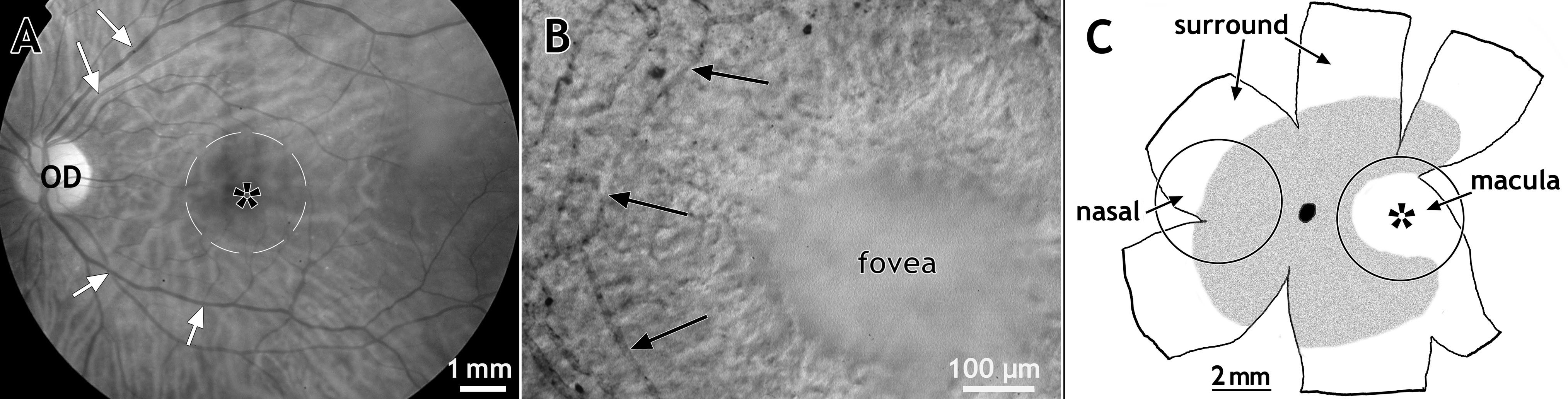
 Figure 1 of Kozulin, Mol Vis 2009; 15:45-59.