Figure 3. Choroidal and scleral muscarinic receptor subtype protein expression in the tree shrew. Tree shrew posterior eye cups were
fixed in 4% paraformaldehyde and reacted with muscarinic monoclonal antibodies to the respective receptor subtypes. A FITC-labeled
secondary antibody was used to visualize the protein distribution in the choroid (CH) and sclera (SC; first column). The corresponding
bright-field section (second column) and negative controls (last column) are also shown. The choroid (CH) and sclera (SC)
are labeled, and the scale bar represents 20 μm.
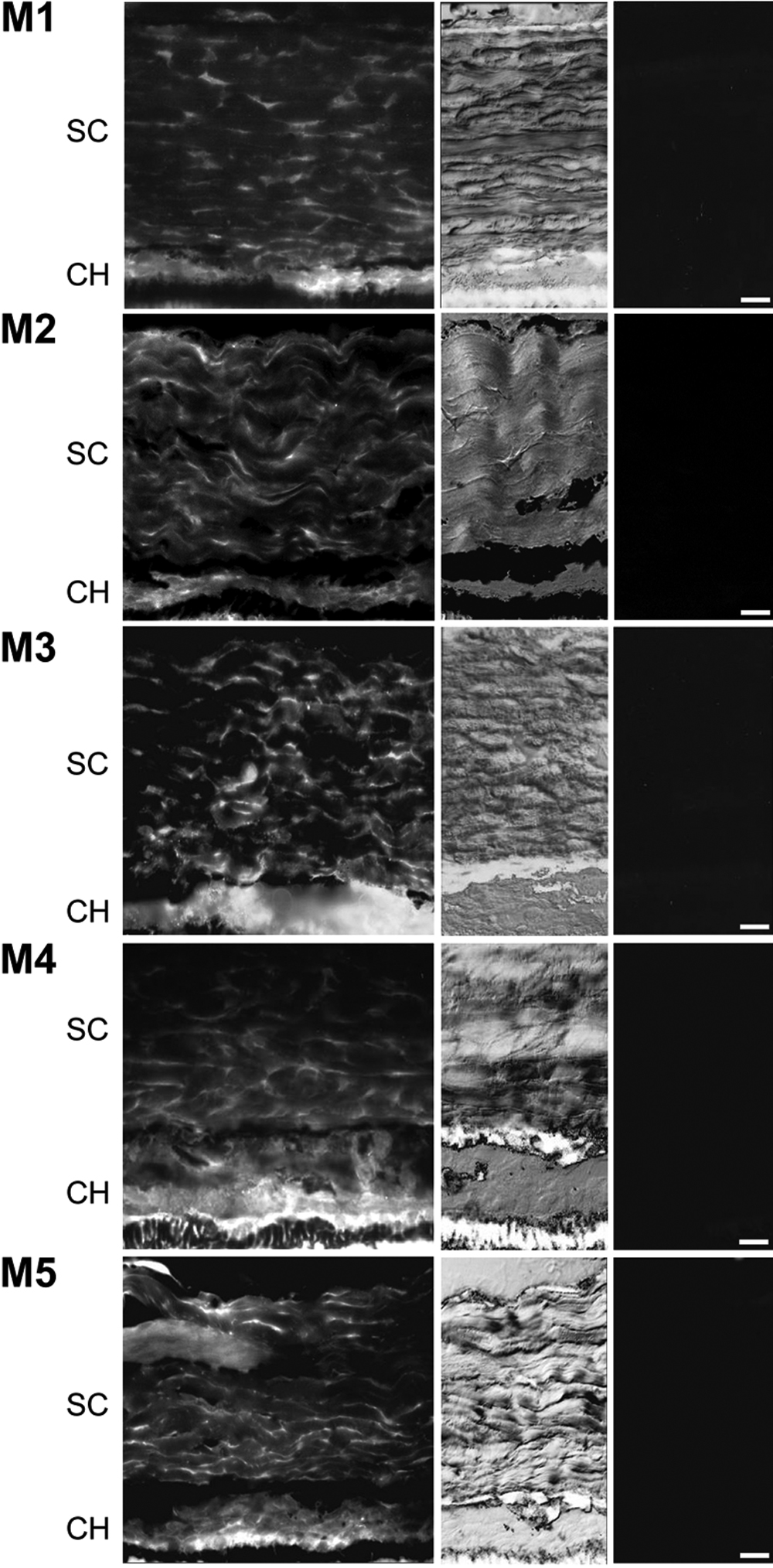
 Figure 3 of
McBrien, Mol Vis 2009; 15:464-475.
Figure 3 of
McBrien, Mol Vis 2009; 15:464-475.