Figure 2. Immunohistochemical staining for
COX-2, and survivin in control sections. Sections from Crohn’s disease (A)
and human cutaneous malignant melanoma (C) were included as
positive controls. No immunostaining for COX-2 (E) and survivin (F)
in normal conjunctiva was observed. Sections incubated without a
primary antibody (inset B) or with an isotype control antibody
(inset D) displayed no immunoreactivity. Each section was
counterstained with hematoxylin. Original magnification, A:
200X; C,E,F: 400X; insets B,D:
400X.
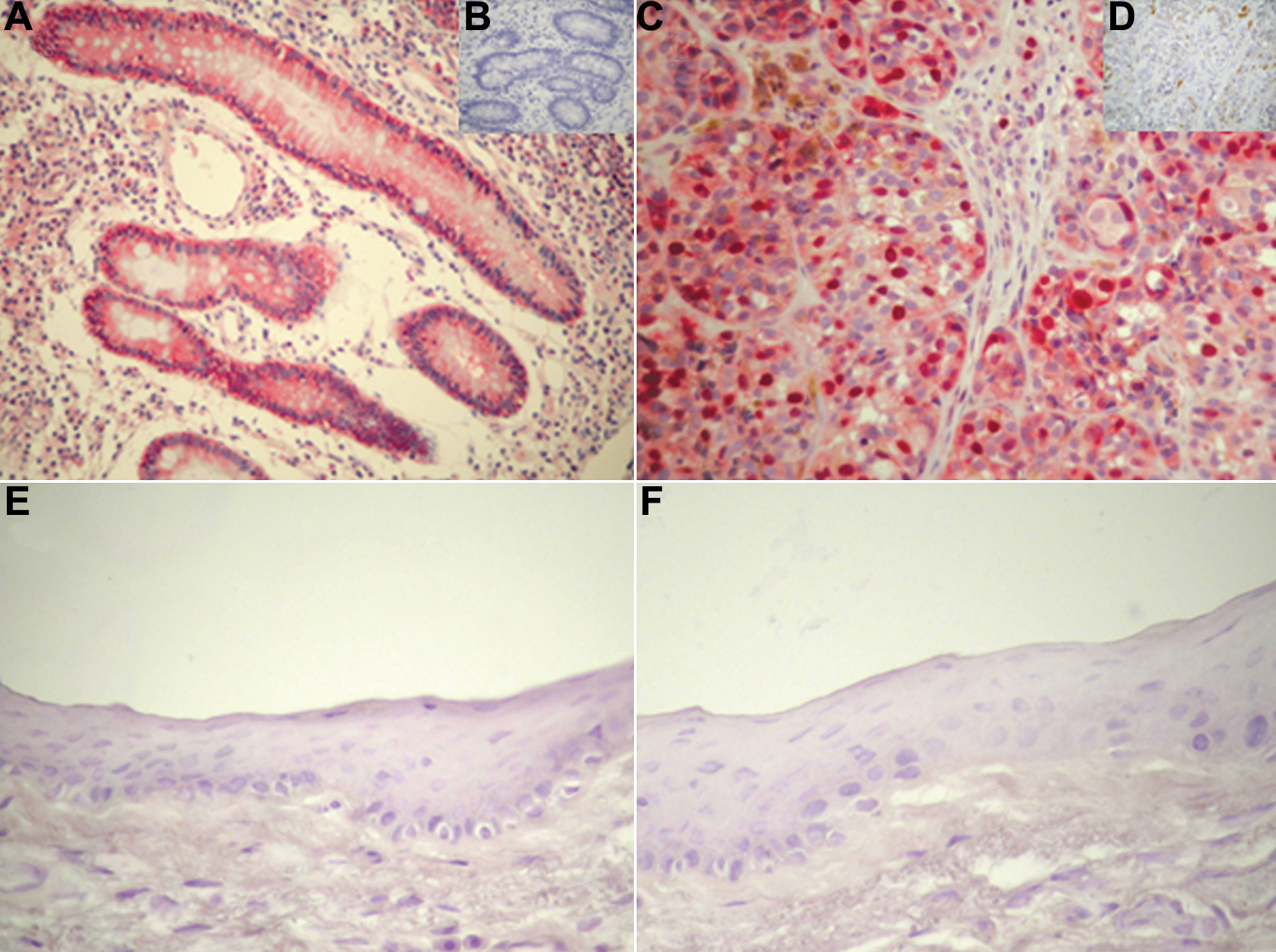
 Figure 2 of Maxia, Mol Vis 2009; 15:458-463.